 Explore
 Validate
 Learn
Learn Immunocytochemistry
Antibody data
- Antibody Data
- Antigen structure
- References [0]
- Comments [0]
- Validations
- Immunocytochemistry [1]
- Immunohistochemistry [7]
Submit
Validation data
Reference
Comment
Report error
- Product number
- AMAb91362 - Provider product page

- Provider
- Atlas Antibodies
- Proper citation
- Atlas Antibodies Cat#AMAb91362, RRID:AB_2716652
- Product name
- Anti-FOXP2
- Antibody type
- Monoclonal
- Reactivity
- Human, Mouse, Rat
- Host
- Mouse
- Conjugate
- Unconjugated
- Antigen sequence
AQQLVFQQQLLQMQQLQQQQHLLSLQRQGLISIPP
GQAALPVQSLPQAGLSPAEIQQLWKEVTGVHSMED
NGIKHGGLDLTTNNSSSTTSSNTSKASPPITHHS- Isotype
- IgG
- Antibody clone number
- CL5312
- Vial size
- 100 µl
- Storage
- Store at +4°C for short term storage. Long time storage is recommended at -20°C.
No comments: Submit comment
Supportive validation
- Submitted by
- Atlas Antibodies (provider)
- Main image

- Experimental details
- Immunofluorescence staining of RH-30 cells using the Anti-FOXP2 monoclonal antibody, showing specific staining in the nucleoplasm in green. Microtubule- and nuclear probes are visualized in red and blue, respectively (where available).
- Sample type
- HUMAN

Supportive validation
- Submitted by
- Atlas Antibodies (provider)
- Main image

- Experimental details
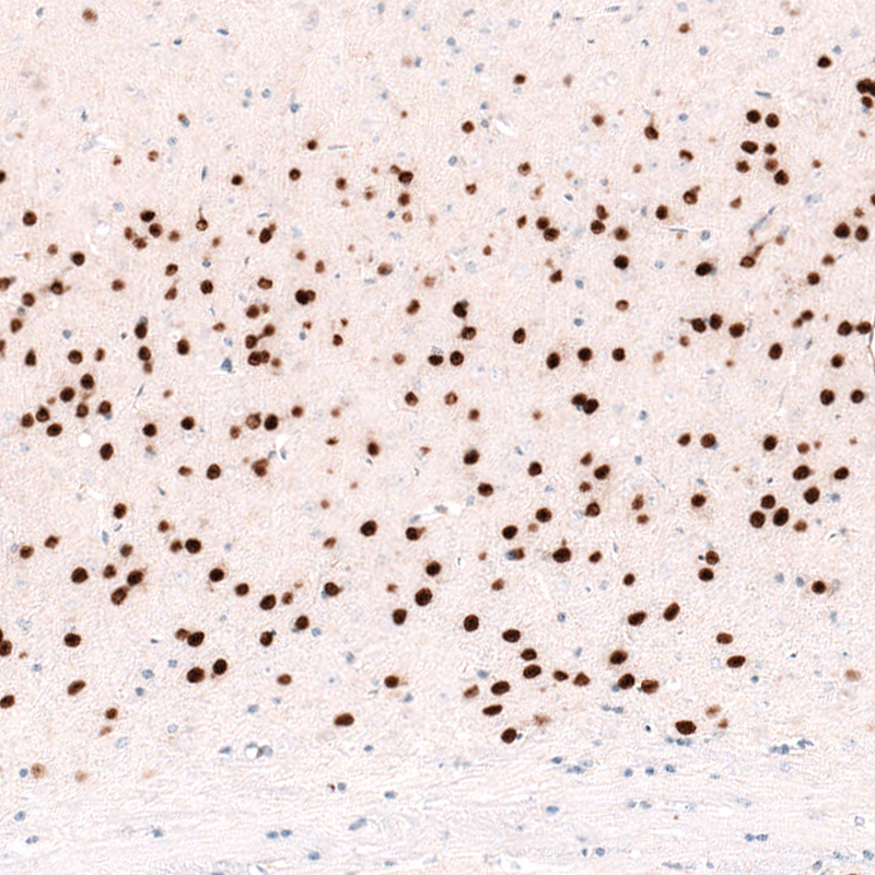
- Immunohistochemical staining of rat brain shows nuclear immunoreactivity in cortical layer 6 neurons, as well as in caudatoputamen.

- Submitted by
- Atlas Antibodies (provider)
- Main image

- Experimental details
- Immunohistochemical staining of rat cerebral cortex shows strong nuclear immunoreactivity in neurons in layer 6.

- Submitted by
- Atlas Antibodies (provider)
- Main image

- Experimental details
- Immunohistochemical staining of mouse brain shows nuclear immunoreactivity in cortical layer 6 neurons, as well as in caudatoputamen.

- Submitted by
- Atlas Antibodies (provider)
- Main image

- Experimental details
- Immunohistochemical staining of mouse cerebral cortex shows strong nuclear immunoreactivity in neurons in layer 6.
- Submitted by
- Atlas Antibodies (provider)
- Main image

- Experimental details
- Immunohistochemical staining of human cerebral cortex shows nuclear immunoreactivity in neurons in layer 6.
- Submitted by
- Atlas Antibodies (provider)
- Main image

- Experimental details
- Immunohistochemical staining of human cerebral cortex shows strong nuclear immunoreactivity in neurons in layer 6.

- Submitted by
- Atlas Antibodies (provider)
- Main image

- Experimental details
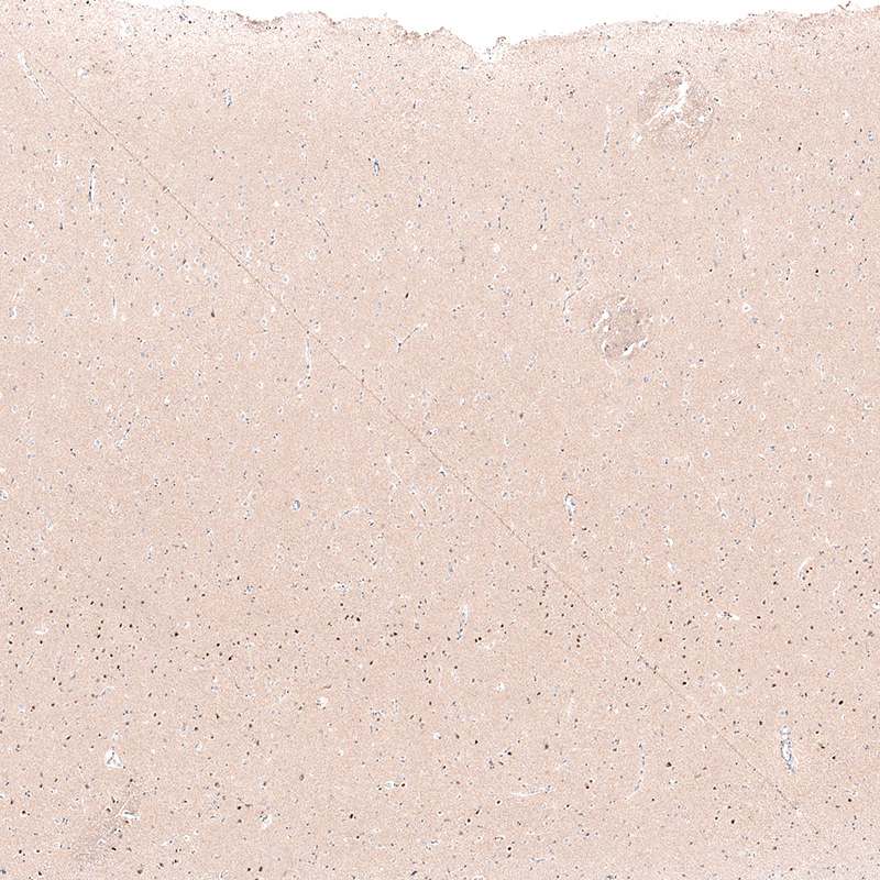
- Immunohistochemical staining of human lymph node shows absence of positivity in lymphoid cells as expected (negative control).
