Explore
 Validate
 Learn
Learn Western blot
 Immunohistochemistry
Antibody data
- Antibody Data
- Antigen structure
- References [2]
- Comments [0]
- Validations
- Immunohistochemistry [2]
- Flow cytometry [4]
- Other assay [2]
Submit
Validation data
Reference
Comment
Report error
- Product number
- PA5-78897 - Provider product page

- Provider
- Invitrogen Antibodies
- Product name
- Carbonic Anhydrase II Polyclonal Antibody
- Antibody type
- Polyclonal
- Antigen
- Recombinant full-length protein
- Description
- Reconstitute with 0.2 mL of distilled water to yield a concentration of 500 µg/mL. Positive Control - WB: human 293T whole cell, human THP-1 whole cell, human HEL whole cell, rat kidney tissue, rat stomach tissue, mouse kidney tissue, mouse stomach tissue. IHC: human gastric cancer tissue, human liver cancer tissue. Flow: HT-29 cell, SW480 cell.
- Reactivity
- Human, Mouse, Rat
- Host
- Rabbit
- Isotype
- IgG
- Vial size
- 100 μg
- Concentration
- 500 μg/mL
- Storage
- -20°C
Submitted references Zinc is a master-regulator of sperm function associated with binding, motility, and metabolic modulation during porcine sperm capacitation.
A Novel Sulfonamide, 4-FS, Reduces Ethanol Drinking and Physical Withdrawal Associated With Ethanol Dependence.
Zigo M, Kerns K, Sen S, Essien C, Oko R, Xu D, Sutovsky P
Communications biology 2022 Jun 3;5(1):538
Communications biology 2022 Jun 3;5(1):538
A Novel Sulfonamide, 4-FS, Reduces Ethanol Drinking and Physical Withdrawal Associated With Ethanol Dependence.
Sona Khan M, Trenet W, Xing N, Sibley B, Abbas M, Al-Rashida M, Rauf K, Mandyam CD
International journal of molecular sciences 2020 Jun 21;21(12)
International journal of molecular sciences 2020 Jun 21;21(12)
No comments: Submit comment
Supportive validation
- Submitted by
- Invitrogen Antibodies (provider)
- Main image
- Experimental details
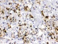
- Immunohistochemistry analysis of Carbonic Anhydrase II on paraffin-embedded human gastric cancer tissue. Antigen retrieval was performed using citrate buffer (pH6, epitope retrieval solution) for 20 mins. Sample was blocked using 10% goat serum, incubated with Carbonic Anhydrase II polyclonal antibody (Product# PA5-78897) with a dilution of 1 µg/mL (overnight at 4°C), and followed by biotinylated goat anti-rabbit IgG (30 minutes at 37°C). Development was performed using Streptavidin-Biotin-Complex (SABC) with DAB chromogen method.
- Submitted by
- Invitrogen Antibodies (provider)
- Main image
- Experimental details
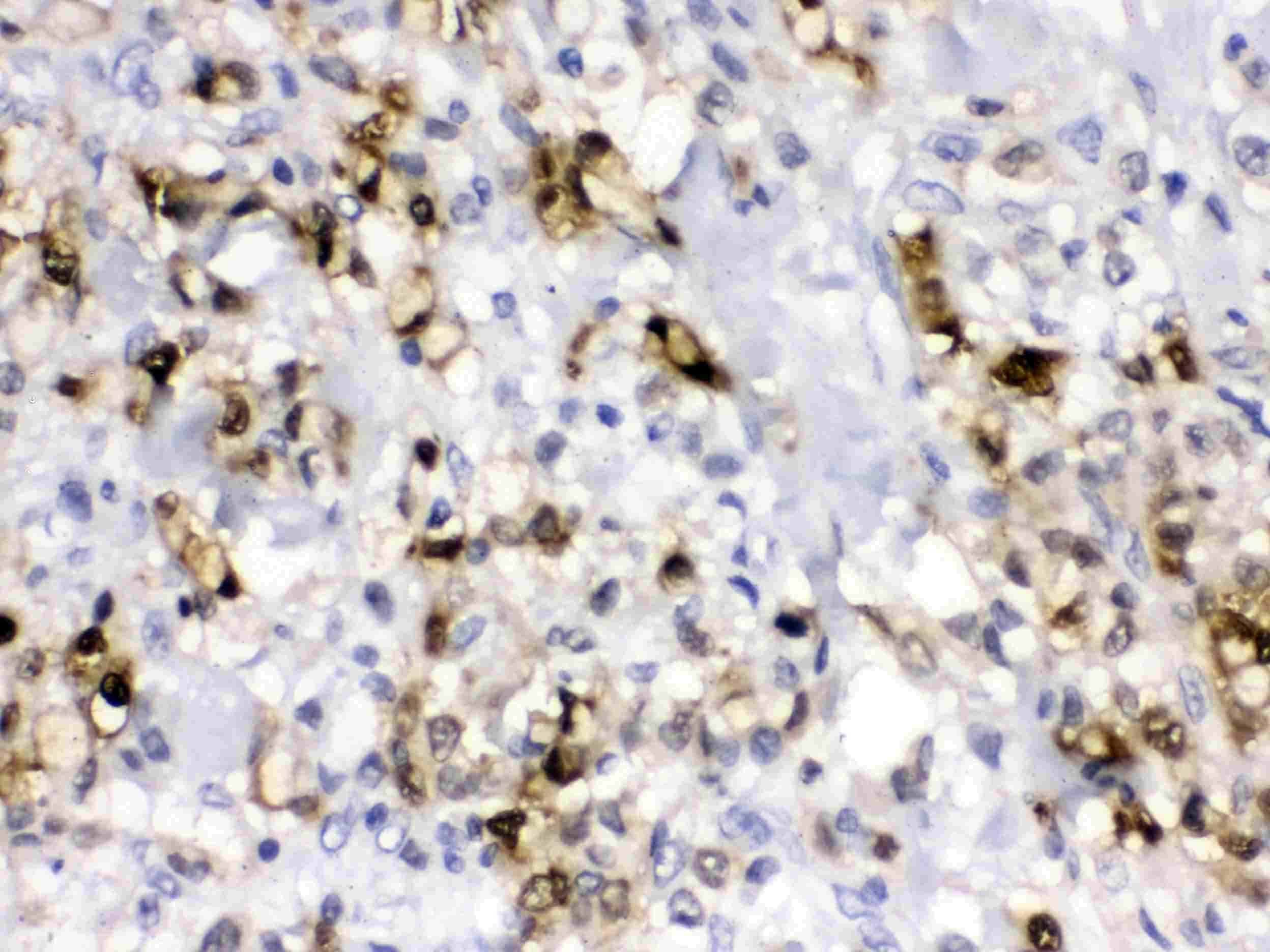
- Immunohistochemistry analysis of Carbonic Anhydrase II on paraffin-embedded human liver cancer tissue. Antigen retrieval was performed using citrate buffer (pH6, epitope retrieval solution) for 20 mins. Sample was blocked using 10% goat serum, incubated with Carbonic Anhydrase II polyclonal antibody (Product# PA5-78897) with a dilution of 1 µg/mL (overnight at 4°C), and followed by biotinylated goat anti-rabbit IgG (30 minutes at 37°C). Development was performed using Streptavidin-Biotin-Complex (SABC) with DAB chromogen method.
Supportive validation
- Submitted by
- Invitrogen Antibodies (provider)
- Main image
- Experimental details
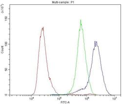
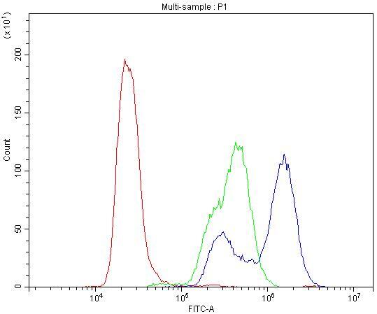
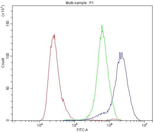
- Flow Cytometry of Carbonic Anhydrase II in HT-29 cells (blue line), isotype control rabbit IgG (green line) and unlabeled (red line). Samples were blocked with 10% goat serum, incubated with Carbonic Anhydrase II Polyclonal Antibody (Product # PA5-78897) at a dilution of 1 μg (per 1x10^6 cells), followed by DyLight®488 conjugated goat anti-rabbit IgG (for 30 minutes at 20°C) using 5-10 μg (per 1x10^6 cells) dilution.

- Submitted by
- Invitrogen Antibodies (provider)
- Main image
- Experimental details
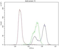
- Flow Cytometry of Carbonic Anhydrase II in SW480 cells (blue line), isotype control rabbit IgG (green line) and unlabeled (red line). Samples were blocked with 10% goat serum, incubated with Carbonic Anhydrase II Polyclonal Antibody (Product # PA5-78897) at a dilution of 1 μg (per 1x10^6 cells), followed by DyLight®488 conjugated goat anti-rabbit IgG (for 30 minutes at 20°C) using 5-10 μg (per 1x10^6 cells) dilution.
- Submitted by
- Invitrogen Antibodies (provider)
- Main image

- Experimental details
- Flow Cytometry of Carbonic Anhydrase II in HT-29 cells (blue line), isotype control rabbit IgG (green line) and unlabeled (red line). Samples were blocked with 10% goat serum, incubated with Carbonic Anhydrase II Polyclonal Antibody (Product # PA5-78897) at a dilution of 1 μg (per 1x10^6 cells), followed by DyLight®488 conjugated goat anti-rabbit IgG (for 30 minutes at 20°C) using 5-10 μg (per 1x10^6 cells) dilution.
- Submitted by
- Invitrogen Antibodies (provider)
- Main image

- Experimental details
- Flow Cytometry of Carbonic Anhydrase II in SW480 cells (blue line), isotype control rabbit IgG (green line) and unlabeled (red line). Samples were blocked with 10% goat serum, incubated with Carbonic Anhydrase II Polyclonal Antibody (Product # PA5-78897) at a dilution of 1 μg (per 1x10^6 cells), followed by DyLight®488 conjugated goat anti-rabbit IgG (for 30 minutes at 20°C) using 5-10 μg (per 1x10^6 cells) dilution.

Supportive validation
- Submitted by
- Invitrogen Antibodies (provider)
- Main image

- Experimental details
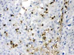
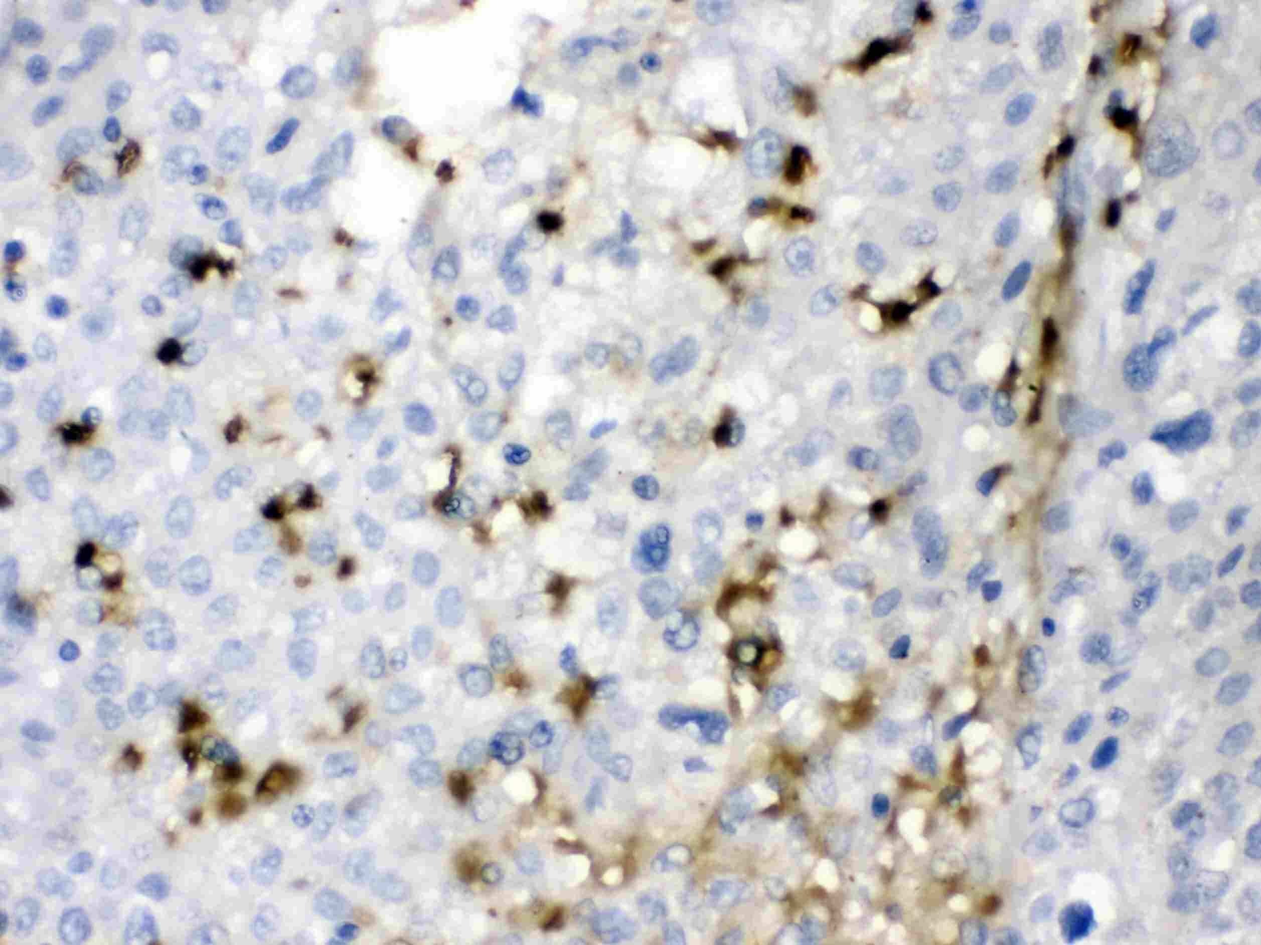
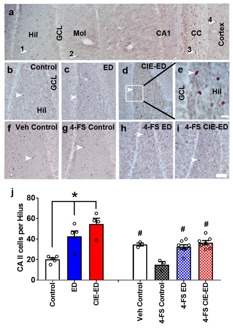
- Figure 3 Carbonic anhydrase type II (CA II) expression in the adult rat hippocampus. ( a ) Photomicrograph of CA II immunohistochemistry in the hippocampus and cortex from one control rat. CA II+ cells appeared as single cells; each immunoreactive cell is pointed with an arrowhead. 1- CA II+ cell in the hilus (Hil); 2-CA II+ cell in the molecular layer (Mol); 3-CA II+ cell in the corpus callosum (cc); 4-CA II+ cell in the cortex. ( b - i ) 100x images of the hilus used for quantitative analyses of CA II cells. ( e ) Zoomed in image shown in ( d ) to indicate the morphology of CA II+ cells in the hilus. Scale bar in ( e ) is 20 um; scale bar in ( i ) is 50 um (applies b - d and f - i ). ( j ) Number of CA II+ cells in the hilus. n = 5 controls, n = 5 ED, n = 4 CIE-ED, n = 3 vehicle controls, n = 3 4-FS controls, n = 8 4-FS ED rats, n = 7 4-FS CIE-ED rats. * p < 0.05, compared to controls; # p < 0.05 compared to 4-FS control. Data are expressed as mean +- S.E.M.
- Submitted by
- Invitrogen Antibodies (provider)
- Main image
- Experimental details
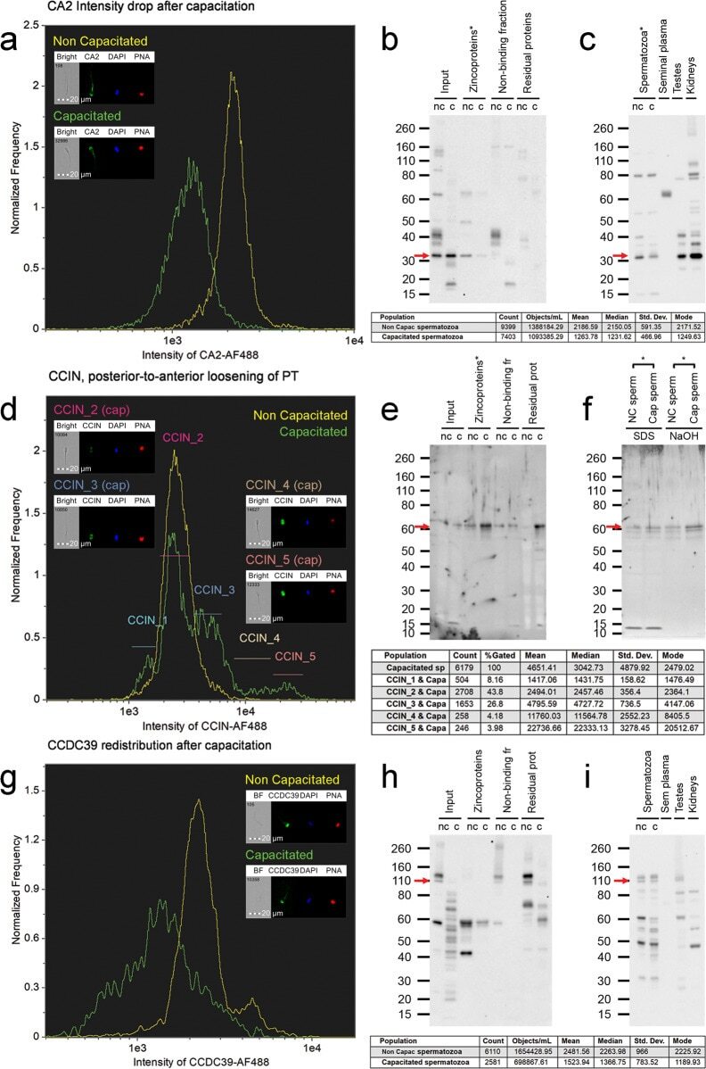
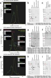
- Phenotyping of three significantly different zincoproteins between non-capacitated and IVC spermatozoa. a - c CA2, d - e CCIN, and g - i CCDC39. IBFC of formaldehyde fixed and Triton X-100 permeabilized ( a , d ) or methanol fixed/permeabilized ( g ) spermatozoa; labeled with corresponding primary antibodies. Appropriate species-specific secondary antibodies conjugated to AF488 were used and coincubated with DAPI nuclear stain and PNA-AF647 for acrosome detection. Negative controls with respective non-immune sera of matching immunoglobulin concentration were performed and were published in our previous study. IBFC was performed in 5 replicates with consistent results. WB detection in individual fractions of zincoproteome purification ( b , e , h ), and WB detection in whole sperm extracts ( c , f , i ) were employed to fully characterize the capacitation changes in the studied proteins. The red arrows point to the bands of actual (CA2 ~ 32 kDa, CCIN ~ 60 kDa), and predicted (CCDC39 ~ 110 kDa) molecular weights.WB detection in sperm zincoproteome ( b , e , h ) and the whole sperm extracts ( c , f , i ) was performed in 8 replicates, and 3 replicates respectively. A statistically significant difference ( P < 0.05, two-sample t -test) between non-capacitated and capacitated spermatozoa is indicated by an asterisk.