Explore
 Validate
 Learn
Learn Western blot
 Immunohistochemistry
Antibody data
- Antibody Data
- Antigen structure
- References [2]
- Comments [0]
- Validations
- Immunohistochemistry [1]
Submit
Validation data
Reference
Comment
Report error
- Product number
- HPA001550 - Provider product page

- Provider
- Atlas Antibodies
- Proper citation
- Atlas Antibodies Cat#HPA001550, RRID:AB_1078393
- Product name
- Anti-CA2
- Antibody type
- Polyclonal
- Description
- Polyclonal Antibody against Human CA2, Gene description: carbonic anhydrase II, Alternative Gene Names: CA-II, CAII, Car2, Validated applications: WB, IHC, Uniprot ID: P00918, Storage: Store at +4°C for short term storage. Long time storage is recommended at -20°C.
- Reactivity
- Human, Mouse, Rat
- Host
- Rabbit
- Conjugate
- Unconjugated
- Isotype
- IgG
- Vial size
- 100 µl
- Concentration
- 0.1 mg/ml
- Storage
- Store at +4°C for short term storage. Long time storage is recommended at -20°C.
- Handling
- The antibody solution should be gently mixed before use.
Submitted references Suspension culture in a rotating bioreactor for efficient generation of human intestinal organoids
Low CA II expression is associated with tumor aggressiveness and poor prognosis in gastric cancer patients.
Takahashi J, Mizutani T, Sugihara H, Nagata S, Kato S, Hiraguri Y, Takeoka S, Tsuchiya M, Kuno R, Kakinuma S, Watanabe M, Okamoto R
Cell Reports Methods 2022;2(11):100337
Cell Reports Methods 2022;2(11):100337
Low CA II expression is associated with tumor aggressiveness and poor prognosis in gastric cancer patients.
Hu X, Huang Z, Liao Z, He C, Fang X
International journal of clinical and experimental pathology 2014;7(10):6716-24
International journal of clinical and experimental pathology 2014;7(10):6716-24
No comments: Submit comment
Supportive validation
- Submitted by
- Atlas Antibodies (provider)
- Enhanced method
- Orthogonal validation
- Main image
- Experimental details
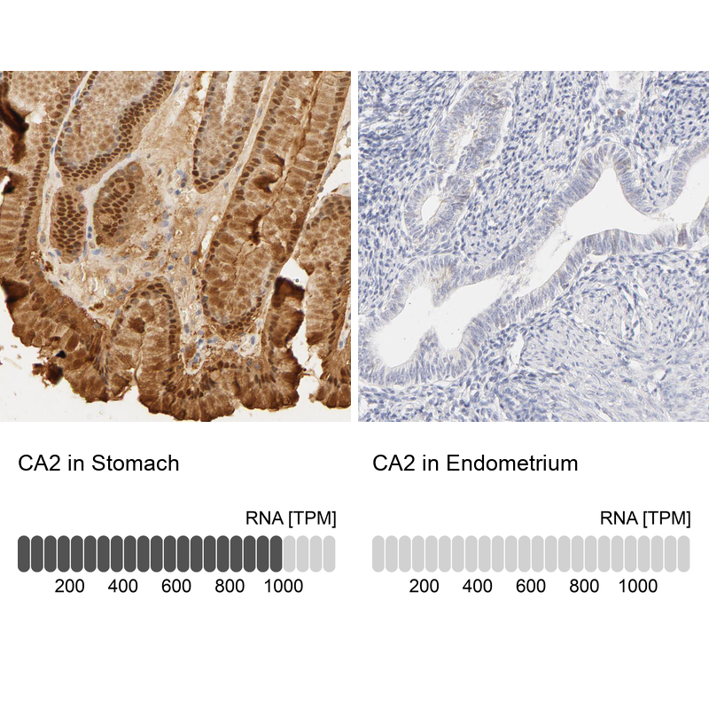
- Immunohistochemistry analysis in human stomach and endometrium tissues using HPA001550 antibody. Corresponding CA2 RNA-seq data are presented for the same tissues.
- Sample type
- Human
- Protocol
- Protocol