Explore
 Validate
 Learn
Learn Western blot
 Immunoprecipitation
 Other assay
Antibody data
- Antibody Data
- Antigen structure
- References [2]
- Comments [0]
- Validations
- Other assay [8]
Submit
Validation data
Reference
Comment
Report error
- Product number
- MA1-24696 - Provider product page

- Provider
- Invitrogen Antibodies
- Product name
- DAPK1 Monoclonal Antibody (DAPK-55)
- Antibody type
- Monoclonal
- Antigen
- Recombinant full-length protein
- Description
- Recommended positive controls: Cos-7. Store product as a concentrated solution. Centrifuge briefly prior to opening the vial.
- Reactivity
- Human
- Host
- Mouse
- Isotype
- IgG
- Antibody clone number
- DAPK-55
- Vial size
- 100 μL
- Concentration
- 7.6 mg/mL
- Storage
- Store at 4°C short term. For long term storage, store at -20°C, avoiding freeze/thaw cycles.
Submitted references A Structurally Conserved RNA Element within SARS-CoV-2 ORF1a RNA and S mRNA Regulates Translation in Response to Viral S Protein-Induced Signaling in Human Lung Cells.
DAPK1 Promotes Extrasynaptic GluN2B Phosphorylation and Striatal Spine Instability in the YAC128 Mouse Model of Huntington Disease.
Basu A, Penumutchu S, Nguyen K, Mbonye U, Tolbert BS, Karn J, Komar AA, Mazumder B
Journal of virology 2022 Jan 26;96(2):e0167821
Journal of virology 2022 Jan 26;96(2):e0167821
DAPK1 Promotes Extrasynaptic GluN2B Phosphorylation and Striatal Spine Instability in the YAC128 Mouse Model of Huntington Disease.
Schmidt ME, Caron NS, Aly AE, Lemarié FL, Dal Cengio L, Ko Y, Lazic N, Anderson L, Nguyen B, Raymond LA, Hayden MR
Frontiers in cellular neuroscience 2020;14:590569
Frontiers in cellular neuroscience 2020;14:590569
No comments: Submit comment
Supportive validation
- Submitted by
- Invitrogen Antibodies (provider)
- Main image
- Experimental details
- NULL
- Submitted by
- Invitrogen Antibodies (provider)
- Main image
- Experimental details
- NULL
- Submitted by
- Invitrogen Antibodies (provider)
- Main image
- Experimental details
- NULL
- Submitted by
- Invitrogen Antibodies (provider)
- Main image

- Experimental details
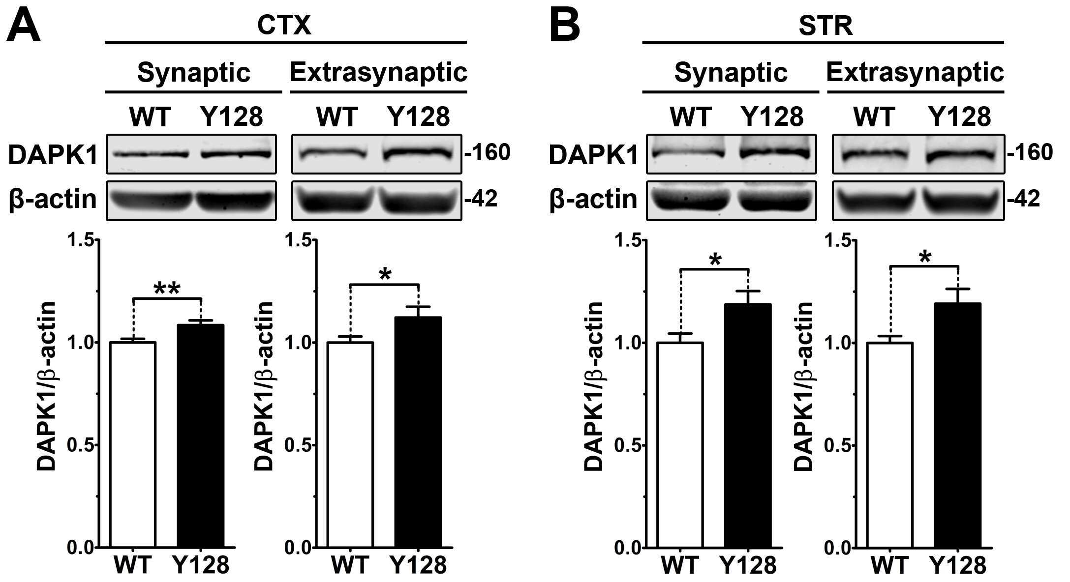
- FIG 8 Treatment of A549 lung cells with SARS-CoV-2 S pseudotyped lentivirus triggers phosphorylation and release of L13a from the ribosome. (a) Detection of ribosome-associated and free L13a in A549 cells exposed to S protein. Extracts from A549 cells treated with ""bald"" (negative control lacking S protein) or S protein pseudotyped lentivirus for the indicated amounts of time were separated into polysome (bottom) and ribosome-free cytosolic (top) fractions, which were then immunoblotted with anti-L13a or anti-L19 antibodies (Thermofisher #14701-1-AP). (b) (left panel), Confirmation of ribosomal and non-ribosomal fractions used in (a). RNA was extracted from the separated fractions using TRIzol, resolved on an agarose gel, and visualized by staining with ethidium bromide. 8b (right panel), Treatment with SARS-CoV-2 S pseudotyped lentivirus induces DAPK-dependent serine phosphorylation of L13a in A549 cells. A549 cells were pretreated with DAPK inhibitor KN62 (or DMSO solvent as a negative control) for 1 h before incubation with the lentivirus. After the indicated amounts of time, cell extracts were prepared and subjected to immunoprecipitation (IP) with anti-L13a antibody, followed by immunoblotting with anti-phosphoserine or anti-L13a antibodies. (c) Requirement of DAPK1 in SARS-CoV-2 S protein-induced and VAIT element-mediated translation control. A549 cells were either untreated or treated with a DAPK1 specific or a non-targeting (used as negative control) siRNA and the s
- Submitted by
- Invitrogen Antibodies (provider)
- Main image
- Experimental details
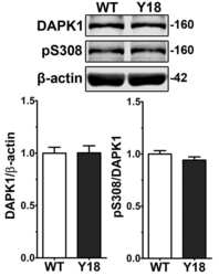
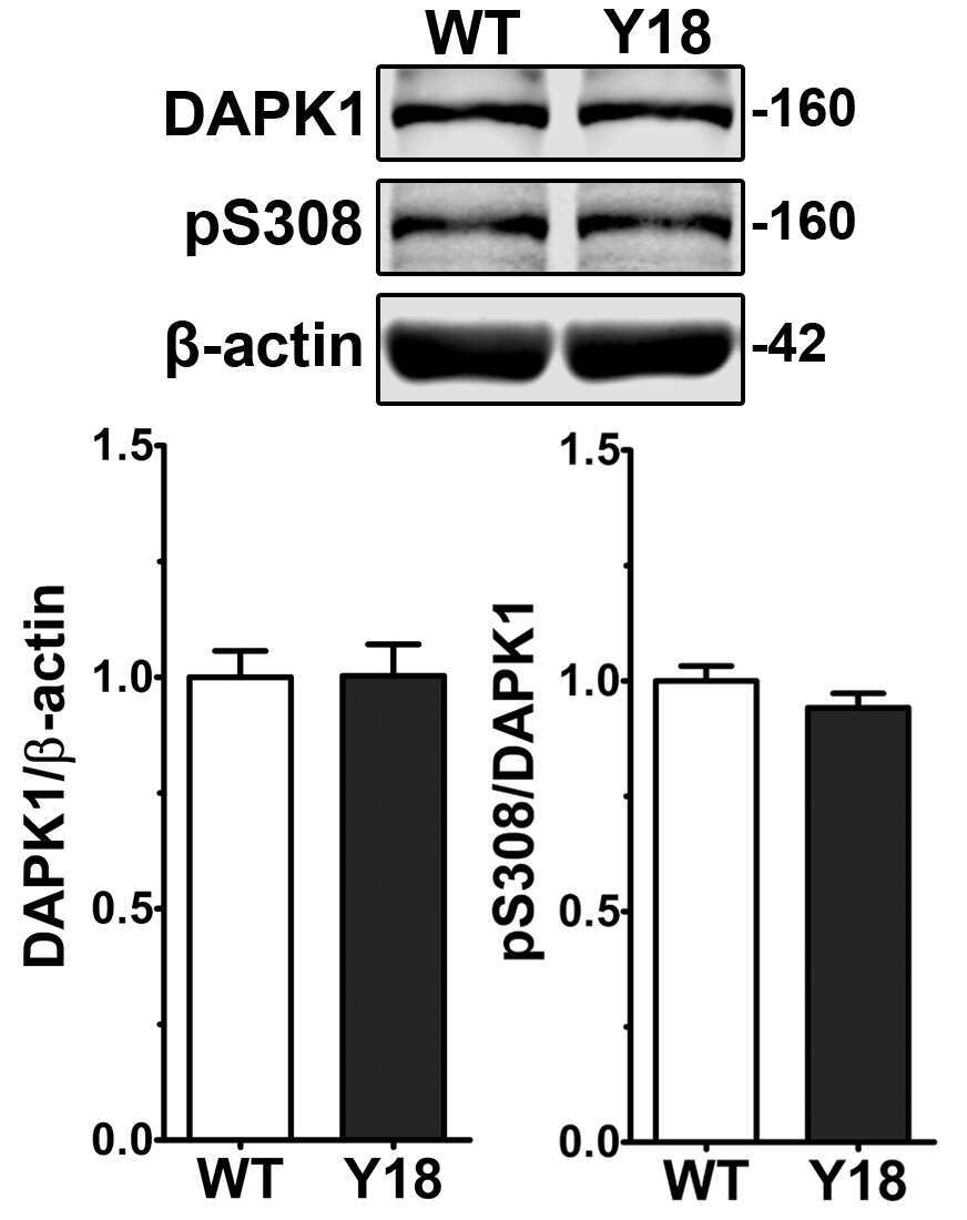
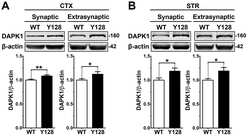
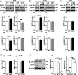
- Figure 1 Increased DAPK1 protein expression and activation occur in affected regions of the YAC128 brain and require mHTT cleavage at D586. (A) Cortical (CTX), (B) striatal (STR), or (C) cerebellar (CB) tissues from 1-month-old WT, YAC128 (Y128; line 53), and C6R (line W13) mice were lysed in a stringent buffer and total lysate was assessed by Western blotting for DAPK1, pS308, and beta-actin protein levels. Data are normalized to WT ( n = 6-12 biological replicates, two technical replicates each; Student's t -test, * p < 0.05, ** p < 0.01, *** p < 0.001). (D) Dapk1 mRNA expression in WT and YAC128 brains at 1 month of age was quantified by qPCR. Data are normalized to WT ( n = 6 biological replicates; Student's t -test). (E) Cortical, striatal, and cerebellar tissues from 1-month-old WT FVB mice were evaluated by Western blot for DAPK1, pS308, and beta-actin protein levels. Data are normalized to CTX values ( n = 8 biological replicates, two technical replicates each; Student's t -test, ### p < 0.001; one-way ANOVA with Bonferroni post hoc analysis, *** p < 0.001).
- Submitted by
- Invitrogen Antibodies (provider)
- Main image
- Experimental details
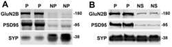
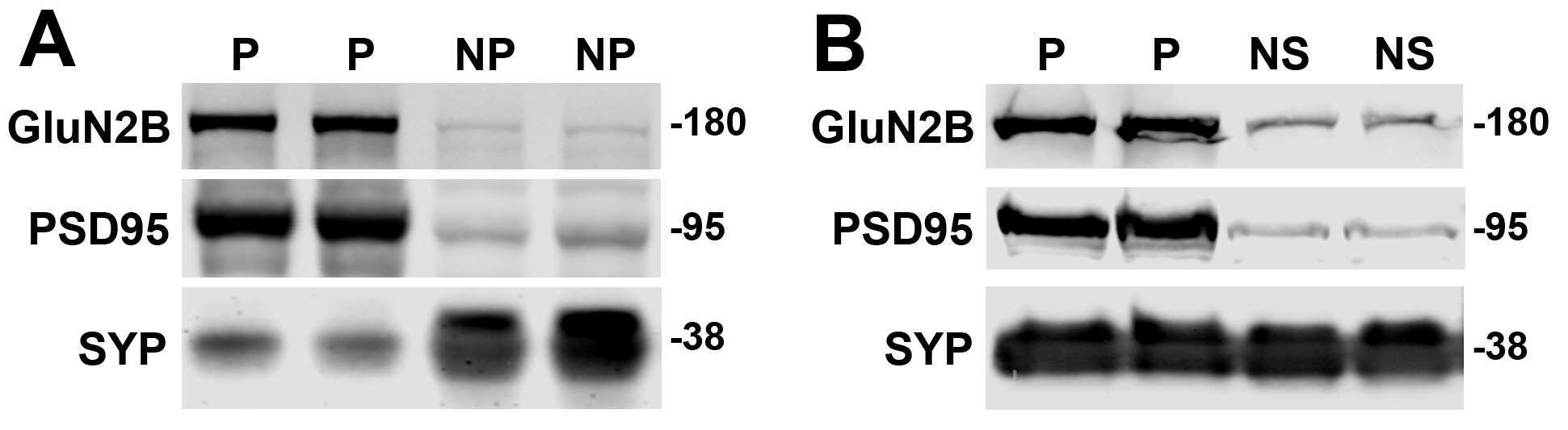
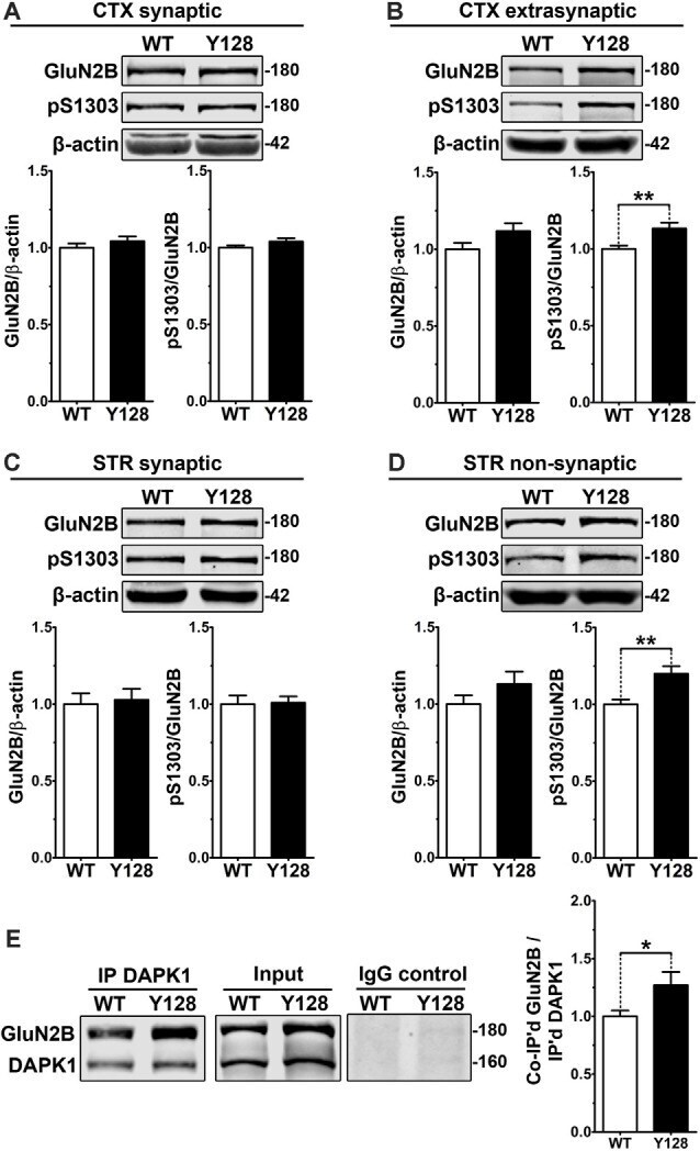
- Figure 2 Extrasynaptic GluN2B S1303 phosphorylation and interaction with DAPK1 are elevated in the YAC128 brain. Cortical or striatal tissues were subjected to subcellular fractionation to yield (A,C) synaptic (PSD), (B) cortical extrasynaptic (non-PSD), or (D) striatal ""non-synaptic"" membrane fractions, which were run by SDS-PAGE and Western blotting for GluN2B expression and S1303 phosphorylation ( n = 6-14 biological replicates, two technical replicates each; Student's t -test, * p < 0.05, ** p < 0.01). (E) DAPK1 was immunoprecipitated from WT and YAC128 cortical lysates, followed by the detection of co-immunoprecipitated GluN2B by Western blot. All sample blot images in panel (E) are cropped from the same Western blot membrane which contained multiple biological replicates run side-by-side. Data are normalized to WT values ( n = 14 biological replicates, Student's t -test, * p < 0.05, ** p < 0.01).
- Submitted by
- Invitrogen Antibodies (provider)
- Main image

- Experimental details
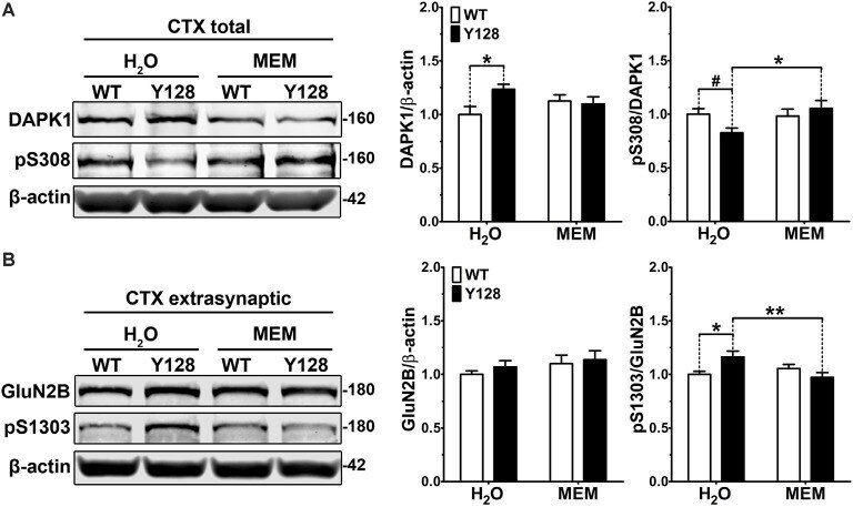
- Figure 3 Low-dose memantine normalizes cortical DAPK1 activation and extrasynaptic pS1303 levels in YAC128 mice. Cortical tissues from 1-month-old WT and YAC128 mice treated with low-dose memantine from conception were processed to obtain (A) total or (B) extrasynaptic (non-PSD) membrane protein fractions. Samples were processed for DAPK1 or GluN2B protein expression and phosphorylation levels by Western blot. Data are normalized to WT H 2 O values ( n = 8 biological replicates, two technical replicates each; two-way ANOVA with Bonferroni post hoc analysis, * p < 0.05, ** p < 0.01; Student's t -test, # p < 0.05; two-way ANOVA interaction * p < 0.05 for DAPK1 protein level and pS308, two-way ANOVA interaction ** p < 0.01 for pS1303).
- Submitted by
- Invitrogen Antibodies (provider)
- Main image
- Experimental details
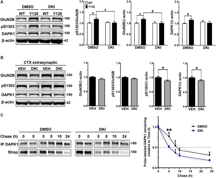
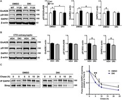
- Figure 4 DAPK1 inhibition normalizes extrasynaptic GluN2B phosphorylation and DAPK1 protein levels in the YAC128 model. (A) Corticostriatal primary cultures from WT and YAC128 embryos were treated at DIV21 with DMSO (0.1%) or a DAPK1 inhibitor (DKI, 10 muM) for 1 h followed by lysis in a gentle buffer to solubilize non-synaptic protein. Lysates were processed by Western blot to detect GluN2B, pS1303, and DAPK1 levels. Data are normalized to WT DMSO values ( n = 8 biological replicates; two-way ANOVA with Bonferroni post hoc analysis, * p < 0.05; Student's t -test, # p < 0.05). (B) Four-week-old YAC128 mice were treated intranasally with 50 nmol of DAPK1 inhibitor (DKI; TC-DAPK6) or vehicle control (VEH). After 6 h, cortical extrasynaptic fractions were isolated to measure GluN2B, pS1303, and DAPK1 levels ( n = 9 biological replicates, two technical replicates each; Student's t -test, * p < 0.05). (C) Quantitative measurement of DAPK1-FLAG protein turnover using bio-orthogonal labeling, FLAG immunoprecipitation, CLICK chemistry, and detection of AHA/Streptavidin (Strep)-labeled DAPK1 in transiently transfected COS-7 cells treated with DMSO or 10 muM DKI (TC-DAPK6) for 24 h ( n = 3 for 8 h and 10 h chase time points, n = 7 for all other chase time points; two-way ANOVA ** p < 0.01 for DKI treatment effect; Sidak's multiple comparisons test, ** p < 0.01).