Explore
 Validate
 Learn
Learn Western blot
 Immunocytochemistry
Antibody data
- Antibody Data
- Antigen structure
- References [3]
- Comments [0]
- Validations
- Immunocytochemistry [1]
Submit
Validation data
Reference
Comment
Report error
- Product number
- HPA001311 - Provider product page

- Provider
- Atlas Antibodies
- Proper citation
- Atlas Antibodies Cat#HPA001311, RRID:AB_1080123
- Product name
- Anti-SUB1
- Antibody type
- Polyclonal
- Description
- Polyclonal Antibody against Human SUB1, Gene description: SUB1 homolog (S. cerevisiae), Alternative Gene Names: p14, p15, PC4, Validated applications: WB, IHC, ICC, Uniprot ID: P53999, Storage: Store at +4°C for short term storage. Long time storage is recommended at -20°C.
- Reactivity
- Human
- Host
- Rabbit
- Conjugate
- Unconjugated
- Isotype
- IgG
- Vial size
- 100 µl
- Concentration
- 0.05 mg/ml
- Storage
- Store at +4°C for short term storage. Long time storage is recommended at -20°C.
- Handling
- The antibody solution should be gently mixed before use.
Submitted references Periodic changes of cyclin D1 mRNA stability are regulated by PC4 modifications in the cell cycle
Single-cell transcriptomic analysis reveals disparate effector differentiation pathways in human Treg compartment
PC4 promotes genome stability and DNA repair through binding of ssDNA at DNA damage sites
Pan Q, Luo P, Hu K, Qiu Y, Liu G, Dai S, Cui B, Yin D, Shi C
Journal of Cell Biology 2024;223(3)
Journal of Cell Biology 2024;223(3)
Single-cell transcriptomic analysis reveals disparate effector differentiation pathways in human Treg compartment
Luo Y, Xu C, Wang B, Niu Q, Su X, Bai Y, Zhu S, Zhao C, Sun Y, Wang J, Liu M, Sun X, Song G, Cui H, Chen X, Huang H, Wang H, Han M, Jiang E, Shi L, Feng X
Nature Communications 2021;12(1)
Nature Communications 2021;12(1)
PC4 promotes genome stability and DNA repair through binding of ssDNA at DNA damage sites
Mortusewicz O, Evers B, Helleday T
Oncogene 2015;35(6):761-770
Oncogene 2015;35(6):761-770
No comments: Submit comment
Supportive validation
- Submitted by
- Atlas Antibodies (provider)
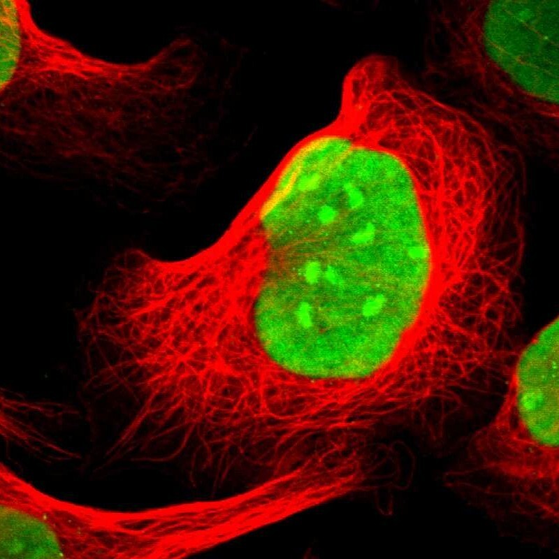
- Main image

- Experimental details
- Immunofluorescent staining of human cell line U-2 OS shows localization to nucleus & nucleoli.
- Sample type
- Human