Explore
 Validate
 Learn
Learn Western blot
Antibody data
- Antibody Data
- Antigen structure
- References [3]
- Comments [0]
- Validations
- Western blot [1]
- Immunocytochemistry [1]
- Immunohistochemistry [1]
- Other assay [4]
Submit
Validation data
Reference
Comment
Report error
- Product number
- PA5-28765 - Provider product page

- Provider
- Invitrogen Antibodies
- Product name
- HUNK Polyclonal Antibody
- Antibody type
- Polyclonal
- Antigen
- Synthetic peptide
- Description
- Recommended positive controls: A431, HeLa. Predicted reactivity: Chimpanzee (100%). Store product as a concentrated solution. Centrifuge briefly prior to opening the vial.
- Reactivity
- Human
- Host
- Rabbit
- Isotype
- IgG
- Vial size
- 100 μL
- Concentration
- 0.65 mg/mL
- Storage
- Store at 4°C short term. For long term storage, store at -20°C, avoiding freeze/thaw cycles.
Submitted references HUNK Phosphorylates Rubicon to Support Autophagy.
Integral membrane protein 2A inhibits cell growth in human breast cancer via enhancing autophagy induction.
Staurosporine, an inhibitor of hormonally up-regulated neu-associated kinase.
Zambrano JN, Eblen ST, Abt M, Rhett JM, Muise-Helmericks R, Yeh ES
International journal of molecular sciences 2019 Nov 19;20(22)
International journal of molecular sciences 2019 Nov 19;20(22)
Integral membrane protein 2A inhibits cell growth in human breast cancer via enhancing autophagy induction.
Zhou C, Wang M, Yang J, Xiong H, Wang Y, Tang J
Cell communication and signaling : CCS 2019 Aug 22;17(1):105
Cell communication and signaling : CCS 2019 Aug 22;17(1):105
Staurosporine, an inhibitor of hormonally up-regulated neu-associated kinase.
Zambrano JN, Williams CJ, Williams CB, Hedgepeth L, Burger P, Dilday T, Eblen ST, Armeson K, Hill EG, Yeh ES
Oncotarget 2018 Nov 13;9(89):35962-35973
Oncotarget 2018 Nov 13;9(89):35962-35973
No comments: Submit comment
Supportive validation
- Submitted by
- Invitrogen Antibodies (provider)
- Main image
- Experimental details
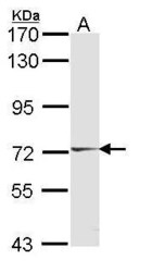
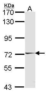
- Western Blot using HUNK Polyclonal Antibody (Product # PA5-28765). Sample (30 µg of whole cell lysate). Lane A: A431 . 7.5% SDS PAGE. HUNK Polyclonal Antibody (Product # PA5-28765) diluted at 1:1,000.
Supportive validation
- Submitted by
- Invitrogen Antibodies (provider)
- Main image
- Experimental details
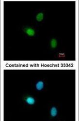
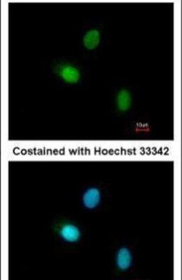
- Immunofluorescent analysis of HUNK in paraformaldehyde-fixed A549 cells using a HUNK polyclonal antibody (Product # PA5-28765) at a 1:200 dilution.
Supportive validation
- Submitted by
- Invitrogen Antibodies (provider)
- Main image

- Experimental details
- Immunohistochemical analysis of paraffin-embedded OVCA xenograft, using HUNK (Product # PA5-28765) antibody at 1:100 dilution. Antigen Retrieval: EDTA based buffer, pH 8.0, 15 min.

Supportive validation
- Submitted by
- Invitrogen Antibodies (provider)
- Main image
- Experimental details
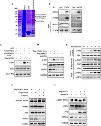
- Fig. 4 ITM2A interacts and is phosphorylated by HUNK. ( a ) Coomassie brilliant blue staining showed that the peptide ""VAIKVIDKKRAKKDTYVTKNLRREGQIQQMI"" existed in the 70 kDa-100 kDa band. ( b ) Co-immunoprecipitation showed the interaction between ITM2A and HUNK in HEK293T cells. ( c ) Flag-HUNK were obtained from the immunoprecipitate in HEK293T cell lysates using Flag antibody. The Flag-HUNK was incubated with the in vitro purified GST-ITM2A WT or T35A protein at 30 degC for 30 min with or without ATP addition, and then the reaction products were subjected to western blotting. ( d ) Flag-ITM2A was obtained from the immunoprecipitate in SKBR-3 cell lysates with or without HUNK knockdown using Flag antibody. Total phosphorylated threonine was detected in the western blotting assay. ( e ) SKBR-3 cells transfected with HA-ITM2A or Flag-HUNK were starved for the indicated times, and HA-ITM2A was immunoprecipitated using HA antibody and used for western blotting analysis. Purified Flag-HUNK was used in the in vitro kinase assay using purified MBP protein as a substrate. ( f ) SKBR-3 cells were transfected with wild-type ITM2A and its T35A mutant simultaneously with or without HUNK siRNA co-transfection for 48 h. Cell lysates were obtained and used for detection by western blotting with the indicated antibodies. ( g ) HEK293T cells were transfected with empty vector and Flag tagged HUNK simultaneously with or without ITM2A siRNA co-transfection for 48 h. Cell lysates were obtain

- Submitted by
- Invitrogen Antibodies (provider)
- Main image
- Experimental details
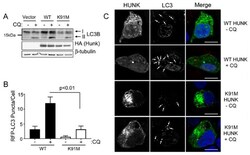
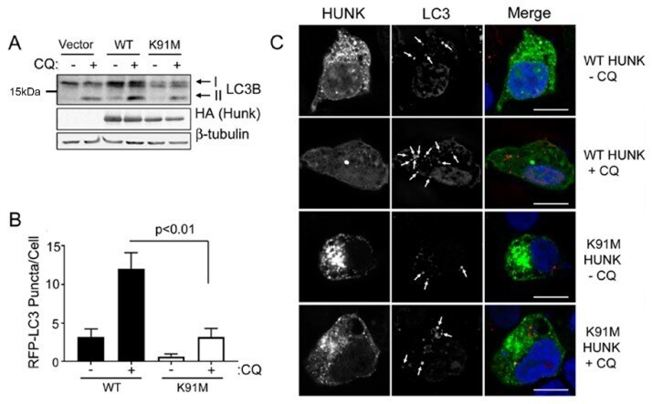
- Figure 1 Hormonally Upregulated Neu-associated Kinase (HUNK) activity is required for HUNK-mediated autophagy. ( A ) 293T cells were transfected with empty vector, HA-HUNK, or HA-K91M HUNK. Cells were then serum deprived and treated with 100 uM CQ for 4 h, lysed and analyzed for LC3B by immunoblot analysis. ( B ) 293T cells plated on coverslips were transfected with RFP-LC3B and either GFP-HUNK or GFP-K91M-HUNK. Cells were serum deprived and treated with 100 uM CQ for 4 h, followed by fixation and imaging. Quantitation of RFP-LC3B puncta in cells. N >= 3 fields per experiment. Data represent 3 or more experiments. Student's T -test was used to perform statistical analysis. ( C ) Representative images of quantitation in ( B ). Scale bar size = 10 um.
- Submitted by
- Invitrogen Antibodies (provider)
- Main image
- Experimental details
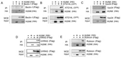
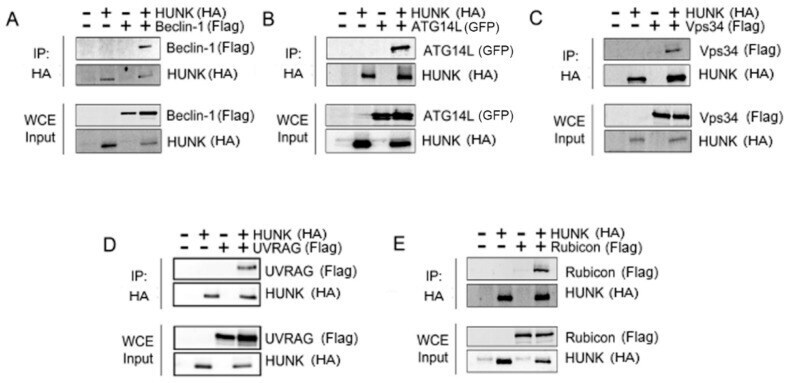
- Figure 2 HUNK binds the Beclin-1 complex members. HA-HUNK and either ( A ) Flag-Beclin-1, ( B ) GFP-Atg14L, ( C ) Flag-Vps34, ( D ) Flag-UVRAG, or ( E ) Flag-Rubicon were co-transfected into 293T cells. HUNK was immunoprecipitated and binding of individual proteins was assessed via immunoblotting for each Beclin-1 complex member. Each member of the Beclin complex co-immunoprecipitated with HUNK. HUNK was immunoprecipitated and detected with anti-HA antibody and Beclin-1 complex members were detected with anti-Flag or anti-GFP antibodies. Interactions were also detected by individual antibodies to each Beclin-1 complex member.
- Submitted by
- Invitrogen Antibodies (provider)
- Main image

- Experimental details
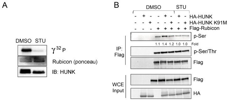
- Figure 3 HUNK phosphorylates Rubicon ( A ) In vitro HUNK kinase assay using recombinant Rubicon (aa 1-375) as substrate. HUNK was preincubated with either DMSO or the HUNK inhibitor staurosporine (STU, 5 uM). ( B ) HA-HUNK or HA-K91M HUNK and Flag-Rubicon were expressed in 293T cells. Flag-Rubicon was immunoprecipitated using anti-Flag affinity resin and isolated protein was immunoblotted using anti-pSer and anti-pSer/Thr antibodies.