 Explore
 Validate
 Learn
Learn Western blot
 Immunocytochemistry
 Immunoprecipitation
Antibody data
- Antibody Data
- Antigen structure
- References [2]
- Comments [0]
- Validations
- Immunocytochemistry [2]
- Other assay [1]
Submit
Validation data
Reference
Comment
Report error
- Product number
- PA5-47907 - Provider product page

- Provider
- Invitrogen Antibodies
- Product name
- RAB27A Polyclonal Antibody
- Antibody type
- Polyclonal
- Antigen
- Recombinant full-length protein
- Description
- This antibody detects human, mouse, and rat RAB27A in Western blots and recombinant human RAB27A in direct ELISAs. In direct ELISAs, less than 1% cross-reactivity with recombinant human RAB27B is observed. Reconstitute in sterile PBS to a final concentration of 0.2 mg/mL.
- Reactivity
- Human, Mouse, Rat
- Host
- Sheep
- Isotype
- IgG
- Vial size
- 100 μg
- Concentration
- 0.2 mg/mL
- Storage
- -20°C, Avoid Freeze/Thaw Cycles
Submitted references Large-scale generation of functional mRNA-encapsulating exosomes via cellular nanoporation.
RF/6A Chorioretinal Cells Do Not Display Key Endothelial Phenotypes.
Yang Z, Shi J, Xie J, Wang Y, Sun J, Liu T, Zhao Y, Zhao X, Wang X, Ma Y, Malkoc V, Chiang C, Deng W, Chen Y, Fu Y, Kwak KJ, Fan Y, Kang C, Yin C, Rhee J, Bertani P, Otero J, Lu W, Yun K, Lee AS, Jiang W, Teng L, Kim BYS, Lee LJ
Nature biomedical engineering 2020 Jan;4(1):69-83
Nature biomedical engineering 2020 Jan;4(1):69-83
RF/6A Chorioretinal Cells Do Not Display Key Endothelial Phenotypes.
Makin RD, Apicella I, Nagasaka Y, Kaneko H, Turner SD, Kerur N, Ambati J, Gelfand BD
Investigative ophthalmology & visual science 2018 Dec 3;59(15):5795-5802
Investigative ophthalmology & visual science 2018 Dec 3;59(15):5795-5802
No comments: Submit comment
Supportive validation
- Submitted by
- Invitrogen Antibodies (provider)
- Main image

- Experimental details
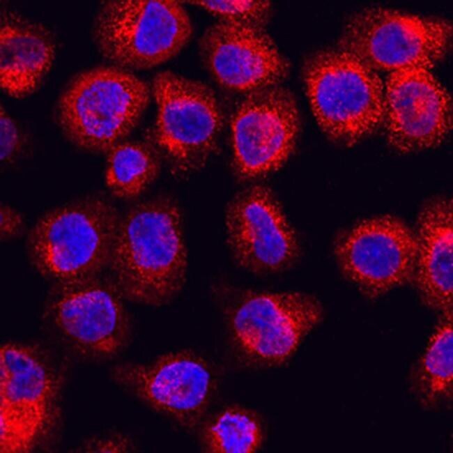
- Immunocytochemical analysis of RAB27A was detected in immersion fixed U937 human histiocytic lymphoma cell line using Sheep Anti-human/mouse/Rat RAB27A Antigen Affinity-purified Polyclonal Antibody (Product # PA5-47907) at 10 µg/mL for 3 hours at room temperature. Cells were stained using the 557-conjugated Anti-Sheep IgG Secondary Antibody (re and counterstained with DAPI (blue). Specific staining was localized to cytoplasm.

- Submitted by
- Invitrogen Antibodies (provider)
- Main image

- Experimental details
- Immunocytochemical analysis of RAB27A was detected in immersion fixed U937 human histiocytic lymphoma cell line using Sheep Anti-human/mouse/Rat RAB27A Antigen Affinity-purified Polyclonal Antibody (Product # PA5-47907) at 10 µg/mL for 3 hours at room temperature. Cells were stained using the 557-conjugated Anti-Sheep IgG Secondary Antibody (re and counterstained with DAPI (blue). Specific staining was localized to cytoplasm.
Supportive validation
- Submitted by
- Invitrogen Antibodies (provider)
- Main image
- Experimental details
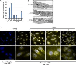
- Figure 2 (A) qPCR analysis of VWF mRNA in HUVEC, HREC, and RF/6A using primers that amplify homologous regions of human and rhesus mRNAs. (B) Transmission electron micrographs of HUVEC, mREC, and RF/6A-1. WPB are long rod-like structures, denoted by W. Scale bar = 2 mum. (C) Low (top) and high (bottom) magnification images of immunofluorescent staining for Rab27a in HUVEC, and RF/6A cells. Note rod-like structures corresponding to WPB in HUVEC, which are absent in RF/6A-1 and -2, and sporadic in RF/6A-3. Scale bar in low magnification = 50 mum, in high magnification = 10 mum.
