 Explore
 Validate
 Learn
Learn Western blot
 Immunoprecipitation
Antibody data
- Antibody Data
- Antigen structure
- References [0]
- Comments [0]
- Validations
- Western blot [5]
- Immunocytochemistry [3]
Submit
Validation data
Reference
Comment
Report error
- Product number
- MA1-23265 - Provider product page

- Provider
- Invitrogen Antibodies
- Product name
- NBS1 Monoclonal Antibody (1D7)
- Antibody type
- Monoclonal
- Antigen
- Other
- Description
- Predicted molecular weight 91 kDa. Positive control: MCF7, HeLa (NE), Raji.
- Reactivity
- Human, Mouse, Rat
- Host
- Mouse
- Isotype
- IgG
- Antibody clone number
- 1D7
- Vial size
- 100 µL
- Concentration
- 1 mg/mL
- Storage
- Store at 4°C short term. For long term storage, store at -20°C, avoiding freeze/thaw cycles.
No comments: Submit comment
Supportive validation
- Submitted by
- Invitrogen Antibodies (provider)
- Main image
- Experimental details
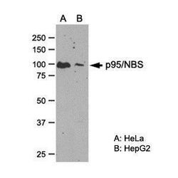
- Western blot analysis of NBS1 in (A) HeLa and (B) HepG2 whole cell extract using a NBS1 monoclonal antibody (Product # MA1-23265).

- Submitted by
- Invitrogen Antibodies (provider)
- Main image

- Experimental details
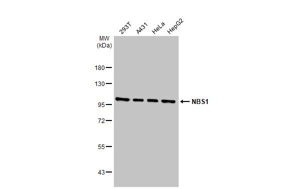
- Western blot analysis of NBS1 in Various whole cell extracts (30 µg). Samples were separated by 7.5% SDS-PAGE and the membrane was probed with NBS1 Monoclonal antibody (Product # MA1-23265) at a dilution of 1:500.

- Submitted by
- Invitrogen Antibodies (provider)
- Main image
- Experimental details
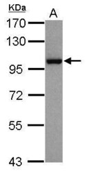
- Western Blot using NBS1 Monoclonal Antibody (1D7) (Product # MA1-23265). Sample (30 µg of whole cell lysate). Lane A: HepG2. 7.5% SDS PAGE. NBS1 Monoclonal Antibody (1D7) (Product # MA1-23265) diluted at 1:1,000. The HRP-conjugated anti-mouse IgG antibody was used to detect the primary antibody.

- Submitted by
- Invitrogen Antibodies (provider)
- Main image

- Experimental details
- Western Blot using NBS1 Monoclonal Antibody (1D7) (Product # MA1-23265). Various whole cell extracts (30 µg) were separated by 7.5% SDS-PAGE, and the membrane was blotted with NBS1 Monoclonal Antibody (1D7) (Product # MA1-23265) diluted at 1:500. The HRP-conjugated anti-mouse IgG antibody was used to detect the primary antibody.
- Submitted by
- Invitrogen Antibodies (provider)
- Main image
- Experimental details
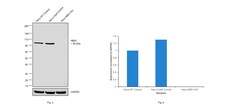
- Knockout of NBS1 was achieved by CRISPR-Cas9 genome editing using LentiArray™ Lentiviral sgRNA (Product # A32042, Assay ID CRISPR791917_LV) and LentiArray Cas9 Lentivirus (Product # A32064). Western blot analysis of NBS1 was performed by loading 30 µg of HeLa Wild Type (Lane 1), HeLa Cas9 (Lane 2) and HeLa NBS1 KO (Lane 3) whole cell extracts. The samples were electrophoresed using NuPAGE™ Novex™ 4-12% Bis-Tris Protein Gel (Product # NP0322BOX). Resolved proteins were then transferred onto a nitrocellulose membrane (Product # IB23001) by iBlot® 2 Dry Blotting System (Product # IB21001). The blot was probed with Anti-NBS1 Monoclonal Antibody (1D7) (Product # MA1-23265, 1:1,000 dilution) and Goat anti-Mouse IgG (H+L) Superclonal™ Recombinant Secondary Antibody, HRP (Product # A28177, 1:4,000 dilution) using the iBright FL 1000 (Product # A32752). Chemiluminescent detection was performed using SuperSignal™ West Dura Extended Duration Substrate (Product # 34076). Loss of signal upon CRISPR mediated knockout (KO) using the LentiArray™ CRISPR product line confirms that antibody is specific to NBS1.

Supportive validation
- Submitted by
- Invitrogen Antibodies (provider)
- Main image
- Experimental details
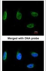
- Immunofluorescent analysis of NBS1 in HeLa cells using a NBS1 monoclonal antibody (Product # MA1-23265) at a 1:100 dilution.

- Submitted by
- Invitrogen Antibodies (provider)
- Main image
- Experimental details
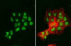
- NBS1 Monoclonal Antibody (1D7) detects NBS1 protein at nucleus by immunofluorescent analysis. Sample: A431 cells were fixed in 4% paraformaldehyde at RT for 15 min. Green: NBS1 stained by NBS1 Monoclonal Antibody (1D7) (Product # MA1-23265) diluted at 1:500. Red: phalloidin, a cytoskeleton marker, diluted at 1:200.

- Submitted by
- Invitrogen Antibodies (provider)
- Main image

- Experimental details
- NBS1 Monoclonal Antibody (1D7) detects NBS1 protein at nucleus by immunofluorescent analysis. Sample: A431 cells were fixed in 4% paraformaldehyde at RT for 15 min. Green: NBS1 stained by NBS1 Monoclonal Antibody (1D7) (Product # MA1-23265) diluted at 1:500. Red: phalloidin, a cytoskeleton marker, diluted at 1:200.
