Explore
 Validate
 Learn
Learn Western blot
Antibody data
- Antibody Data
- Antigen structure
- References [0]
- Comments [0]
- Validations
- Western blot [3]
- Immunohistochemistry [2]
Submit
Validation data
Reference
Comment
Report error
- Product number
- PA3-16528 - Provider product page

- Provider
- Invitrogen Antibodies
- Product name
- NBS1 Polyclonal Antibody
- Antibody type
- Polyclonal
- Antigen
- Other
- Description
- ICC/IF has been performed using methanol-fixed IMR90 primary human fibroblasts.
- Concentration
- Conc. Not Determined
No comments: Submit comment
Supportive validation
- Submitted by
- Invitrogen Antibodies (provider)
- Main image

- Experimental details
- Western blot analysis of NBS1 in human cell lysate using a polyclonal antibody (Product # PA3-16528).

- Submitted by
- Invitrogen Antibodies (provider)
- Main image

- Experimental details
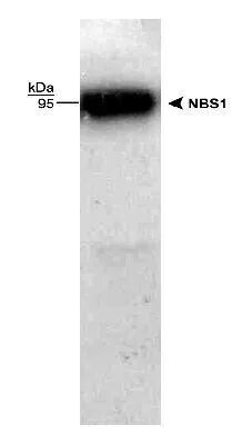
- Knockout validation by Western blot analysis of NBS1 in lysates of HeLa human cervical epithelial carcinoma parental cell line and Nbs1 knockout (KO) HeLa cell line. Samples were incubated in NBS1 polyclonal antibody (Product # PA3-16528) using a dilution of 1:1000 followed by a HRP-conjugated Anti-Rabbit IgG secondary antibody. Specific band was detected for Nbs1 at approximately 95 kDa (as indicated) in the parental HeLa cell line, but is not detectable in the knockout HeLa cell line. This experiment was conducted under reducing conditions.

- Submitted by
- Invitrogen Antibodies (provider)
- Main image

- Experimental details
- Western blot analysis of NBS1 in HeLa whole cell lysate. Samples were incubated in NBS1 polyclonal antibody (Product # PA3-16528). Observed Molecular Weight at ~95 kDa.
Supportive validation
- Submitted by
- Invitrogen Antibodies (provider)
- Main image

- Experimental details
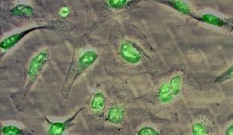
- ICC staining of Hela cells using Product # PA3-16528 anti-NBS1.
- Submitted by
- Invitrogen Antibodies (provider)
- Main image

- Experimental details
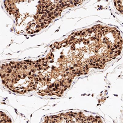
- Immunohistochemical analysis of NBS1 in formalin-fixed paraffin-embedded human testis. Samples were incubated in NBS1 polyclonal antibody (Product # PA3-16528) using a dilution of 1:200. Bond Rx autostainer (Leica Biosystems). The assay involved 20 minutes of heat induced antigen retrieval (HIER) using 10 mM sodium citrate buffer (pH 6.0) and endogenous peroxidase quenching with peroxide block. The sections were incubated with primary antibody for 30 minutes and Bond Polymer Refine Detection (Leica Biosystems) with DAB was used for signal development followed by counterstaining with hematoxylin. Whole slide scanning and capturing of representative images was performed using Aperio AT2 (Leica Biosystems). Staining was performed by Histowiz.