 Explore
 Validate
 Learn
Learn Western blot
Antibody data
- Antibody Data
- Antigen structure
- References [0]
- Comments [0]
- Validations
- Western blot [3]
- Immunocytochemistry [1]
- Immunohistochemistry [1]
Submit
Validation data
Reference
Comment
Report error
- Product number
- PA5-120608 - Provider product page

- Provider
- Invitrogen Antibodies
- Product name
- PTPN2 Polyclonal Antibody
- Antibody type
- Polyclonal
- Antigen
- Recombinant full-length protein
- Description
- Positive test controls include: SW480, BT-474, Jurkat, K-562. The target is usually found in the following locations: Cell membrane, Cytoplasm, Endoplasmic reticulum, Endoplasmic reticulum-Golgi intermediate compartment, Nucleus.
- Reactivity
- Human, Mouse, Rat
- Host
- Rabbit
- Isotype
- IgG
- Vial size
- 100 µL
- Concentration
- 2.48 mg/mL
- Storage
- -20° C, Avoid Freeze/Thaw Cycles
No comments: Submit comment
Supportive validation
- Submitted by
- Invitrogen Antibodies (provider)
- Main image

- Experimental details
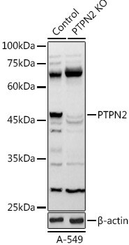
- Western Blot analysis of PTPN2 in HeLa cells using a PTPN2 Polyclonal antibody (Product # PA5-120608) at a dilution of 1:1,000. 25 µg of lysate or protein were loaded into each lane. A blocking buffer of 3% nonfat dry milk in TBST was used. An HRP conjugated Goat Anti-Rabbit IgG (H+L) secondary antibody was used at a 1:10,000 dilution. Chemiluminescent detection was performed for 3s.
- Submitted by
- Invitrogen Antibodies (provider)
- Main image
- Experimental details
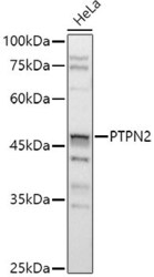
- Western Blot analysis of PTPN2 in HeLa cells using a PTPN2 Polyclonal antibody (Product # PA5-120608) at a dilution of 1:1,000. 25 µg of lysate or protein were loaded into each lane. A blocking buffer of 3% nonfat dry milk in TBST was used. An HRP conjugated Goat Anti-Rabbit IgG (H+L) secondary antibody was used at a 1:10,000 dilution. Chemiluminescent detection was performed for 3s.

- Submitted by
- Invitrogen Antibodies (provider)
- Main image

- Experimental details
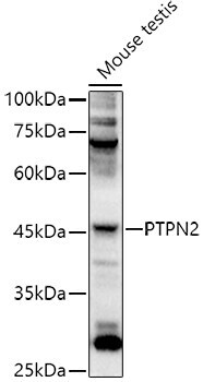
- Western Blot analysis of PTPN2 in HeLa cells using a PTPN2 Polyclonal antibody (Product # PA5-120608) at a dilution of 1:1,000. 25 µg of lysate or protein were loaded into each lane. A blocking buffer of 3% nonfat dry milk in TBST was used. An HRP conjugated Goat Anti-Rabbit IgG (H+L) secondary antibody was used at a 1:10,000 dilution. Chemiluminescent detection was performed for 3s.
Supportive validation
- Submitted by
- Invitrogen Antibodies (provider)
- Main image
- Experimental details
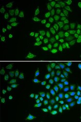
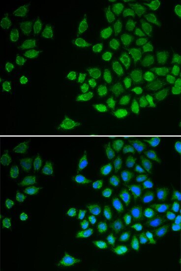
- Immunocytochemical analysis of PTPN2 in MCF-7 cells using a PTPN2 Polyclonal antibody (Product # PA5-120608). Blue: DAPI for nuclear staining.
Supportive validation
- Submitted by
- Invitrogen Antibodies (provider)
- Main image
- Experimental details
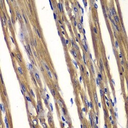
- Immunohistochemical analysis of PTPN2 in rat heart using a Polyclonal antibody (Product # PA5-120608) at a dilution of 1:100.
