Explore
 Validate
 Learn
Learn Western blot
 ELISA
ELISA Immunohistochemistry
Antibody data
- Antibody Data
- Antigen structure
- References [3]
- Comments [0]
- Validations
- Immunohistochemistry [2]
- Flow cytometry [2]
- Other assay [9]
Submit
Validation data
Reference
Comment
Report error
- Product number
- MA5-15883 - Provider product page

- Provider
- Invitrogen Antibodies
- Product name
- IRAK4 Monoclonal Antibody (2H9)
- Antibody type
- Monoclonal
- Antigen
- Purifed from natural sources
- Description
- MA5-15883 targets IRAK4 in indirect ELISA, FACS, IHC, and WB applications and shows reactivity with Human, mouse, and Non-human primate samples. The MA5-15883 immunogen is purified recombinant fragment of human IRAK4 expressed in E. Coli. . MA5-15883 detects IRAK4 which has a predicted molecular weight of approximately 52kDa.
- Reactivity
- Human, Mouse
- Host
- Mouse
- Isotype
- IgG
- Antibody clone number
- 2H9
- Vial size
- 100 μL
- Concentration
- Conc. Not Determined
- Storage
- Store at 4°C short term. For long term storage, store at -20°C, avoiding freeze/thaw cycles.
Submitted references Unexpected Activities in Regulating Ciliation Contribute to Off-target Effects of Targeted Drugs.
Interleukin-1 receptor-associated kinase 4 (IRAK4) plays a dual role in myddosome formation and Toll-like receptor signaling.
Comprehensive RNAi-based screening of human and mouse TLR pathways identifies species-specific preferences in signaling protein use.
Kiseleva AA, Korobeynikov VA, Nikonova AS, Zhang P, Makhov P, Deneka AY, Einarson MB, Serebriiskii IG, Liu H, Peterson JR, Golemis EA
Clinical cancer research : an official journal of the American Association for Cancer Research 2019 Jul 1;25(13):4179-4193
Clinical cancer research : an official journal of the American Association for Cancer Research 2019 Jul 1;25(13):4179-4193
Interleukin-1 receptor-associated kinase 4 (IRAK4) plays a dual role in myddosome formation and Toll-like receptor signaling.
De Nardo D, Balka KR, Cardona Gloria Y, Rao VR, Latz E, Masters SL
The Journal of biological chemistry 2018 Sep 28;293(39):15195-15207
The Journal of biological chemistry 2018 Sep 28;293(39):15195-15207
Comprehensive RNAi-based screening of human and mouse TLR pathways identifies species-specific preferences in signaling protein use.
Sun J, Li N, Oh KS, Dutta B, Vayttaden SJ, Lin B, Ebert TS, De Nardo D, Davis J, Bagirzadeh R, Lounsbury NW, Pasare C, Latz E, Hornung V, Fraser ID
Science signaling 2016 Jan 5;9(409):ra3
Science signaling 2016 Jan 5;9(409):ra3
No comments: Submit comment
Supportive validation
- Submitted by
- Invitrogen Antibodies (provider)
- Main image
- Experimental details
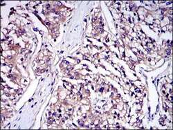
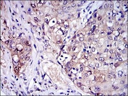
- Immunohistochemical analysis of paraffin-embedded human kidney cancer tissues using IRAK4 monoclonal antibody (Product # MA5-15883) followed with DAB staining.
- Submitted by
- Invitrogen Antibodies (provider)
- Main image
- Experimental details
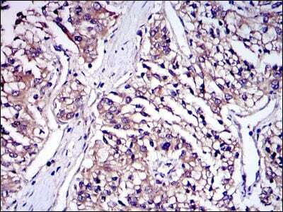
- Immunohistochemical analysis of paraffin-embedded human lung cancer tissues using IRAK4 monoclonal antibody (Product # MA5-15883) followed with DAB staining.

Supportive validation
- Submitted by
- Invitrogen Antibodies (provider)
- Main image
- Experimental details
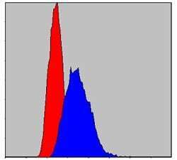
- Flow cytometric analysis of HeLa cells using IRAK4 monoclonal antibody (Product # MA5-15883) (blue) and negative control (red).

- Submitted by
- Invitrogen Antibodies (provider)
- Main image

- Experimental details
- Flow cytometric analysis of HeLa cells using IRAK4 monoclonal antibody (Product # MA5-15883) (blue) and negative control (red).

Supportive validation
- Submitted by
- Invitrogen Antibodies (provider)
- Main image
- Experimental details
- NULL

- Submitted by
- Invitrogen Antibodies (provider)
- Main image
- Experimental details
- NULL
- Submitted by
- Invitrogen Antibodies (provider)
- Main image

- Experimental details
- NULL
- Submitted by
- Invitrogen Antibodies (provider)
- Main image
- Experimental details
- NULL
- Submitted by
- Invitrogen Antibodies (provider)
- Main image
- Experimental details
- NULL
- Submitted by
- Invitrogen Antibodies (provider)
- Main image
- Experimental details
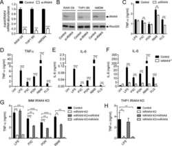
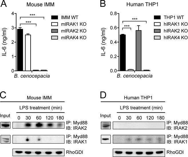
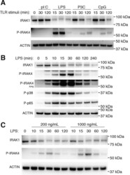
- Figure 1. MyD88-dependent TLR signaling activates IRAK1 and IRAK4. A , BMDMs were left untreated or stimulated with a panel of synthetic TLR ligands for up to 2 h: 10 mug/ml poly(I:C) (pI:C-TLR3); 1 mug/ml LPS (TLR4); 200 ng/ml Pam3CysK4 (P3C-TLR1/2); or 0.1 mu m CpG DNA 1826 (TLR9). WCLs were subjected to immunoblotting for IRAK1, P-IRAK4, and actin as a loading control. The data presented are representative of three independent experiments. B , BMDMs were left untreated or stimulated with 1 mug/ml LPS from 5 min up to 4 h. WCLs were subjected to immunoblotting for IRAK1, P-IRAK4, P-p38 MAPK, NF-kappaB P-p65, and actin as a loading control. The data presented are representative of two independent experiments. C , BMDMs were left untreated or stimulated with either 200 ng/ml or 1 mug/ml LPS from 10 min up to 2 h. WCLs were subjected to immunoblotting for IRAK1, P-IRAK4, and actin as a loading control. The data presented are representative of three independent experiments.

- Submitted by
- Invitrogen Antibodies (provider)
- Main image
- Experimental details
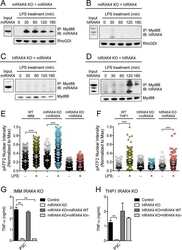
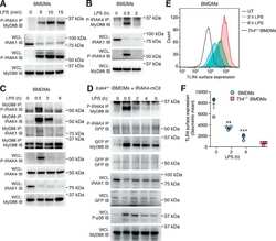
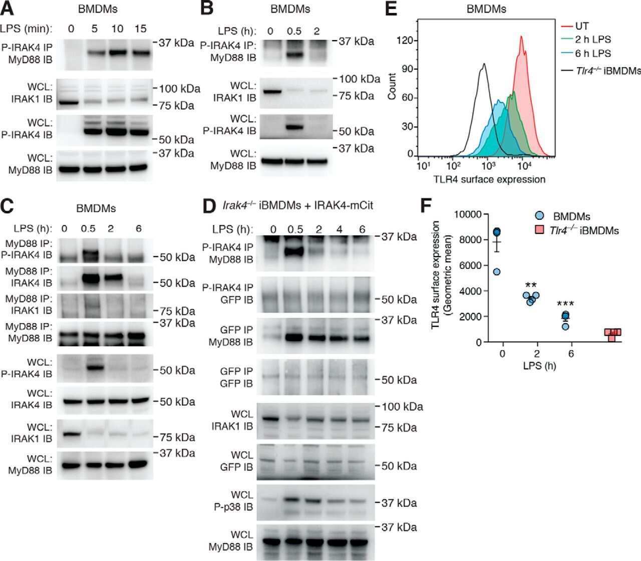
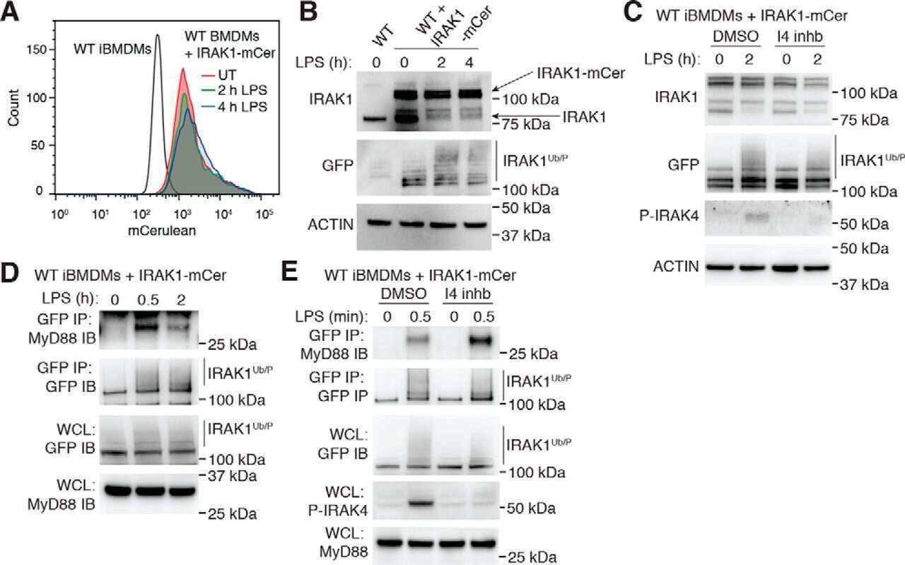
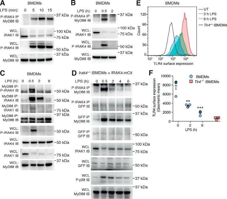
- Figure 2. Myddosome complexes form rapidly but are stable following the loss of P-IRAK4. BMDMs were left untreated or stimulated with 1 mug/ml LPS for 5, 10, and 15 min ( A ) or 30 min and 2 h ( B ). A and B , WCLs were then subjected to P-IRAK4 IP followed by MyD88 immunoblotting or directly subjected to immunoblotting for IRAK-1, P-IRAK4, and MyD88, which also served as a loading control. The data presented are representative of three independent experiments. C , BMDMs were left untreated or stimulated with 1 mug/ml LPS for 30 min, 2 h, or 6 h. WCLs were then subjected to MyD88 IP, followed by immunoblotting for P-IRAK4, IRAK4, IRAK1, and MyD88, or directly subjected to immunoblotting with the same antibodies. The data presented are representative of three independent experiments. D , Irak4 -/- iBMDMs expressing IRAK4-mCitrine were generated as described under ""Experimental procedures."" These cells were left untreated or stimulated with 1 mug/ml LPS for 30 min, 2 h, 4 h, or 6 h before WCLs were divided evenly and subjected to either P-IRAK4 IP or GFP IP followed by immunoblotting for MyD88 and GFP. WCLs were also directly subjected to immunoblotting for IRAK1, GFP, P-p38 MAPK, and MyD88. The data presented are representative of two independent experiments. E , BMDMs were left untreated ( UT ) or stimulated with 1 mug/ml LPS for 2 or 6 h before staining with an antibody against the extracellular region of TLR4 and subjected to flow cytometric analysis to determine
- Submitted by
- Invitrogen Antibodies (provider)
- Main image
- Experimental details
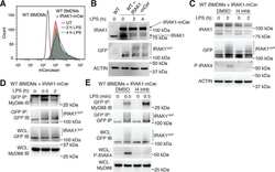
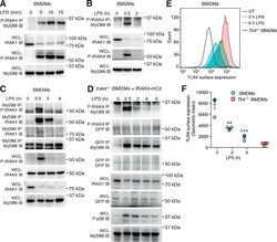
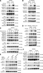
- Figure 4. IRAK4 kinase inhibition stabilizes TLR-induced myddosome interactions. A , BMDMs were pretreated with either DMSO or 20 mu m I4 inhb for 30 min. Cells were then left untreated or stimulated with 1 mug/ml LPS for a further 30 min. WCLs were then subjected to MyD88 IP followed by immunoblotting for IRAK4, IRAK1, P-IRAK4, and MyD88 or directly subjected to immunoblotting with the same antibodies. MyD88 immunoblotting also acted as a loading control. The data presented are representative of three independent experiments. B , WT iBMDMs were treated as in A , before WCLs were subjected to MyD88 IP followed by immunoblotting for IRAK4, IRAK1, and MyD88 or directly subjected to immunoblotting with the same antibodies. The data presented are representative of three independent experiments. C , BMDMs were pretreated with either DMSO or 20 mu m I4 inhb for 30 min. Cells were then left untreated or stimulated with either 500 ng/ml P3C or 0.5 mu m CpG DNA 1826 for a further 30 min. WCLs were then subjected to MyD88 IP followed by immunoblotting for IRAK4, IRAK1, P-IRAK4, and MyD88 or directly subjected to immunoblotting with the same antibodies. MyD88 immunoblotting also acted as a loading control. The data presented are representative of three independent experiments. D , Irak4 -/- iBMDMs expressing IRAK4-mCitrine were treated as in A before WCLs were subjected to GFP IP, followed by immunoblotting for MyD88 and GFP, or directly subjected to immunoblotting for P-IRA
- Submitted by
- Invitrogen Antibodies (provider)
- Main image
- Experimental details
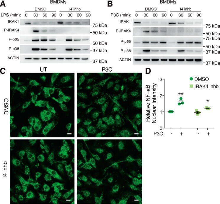
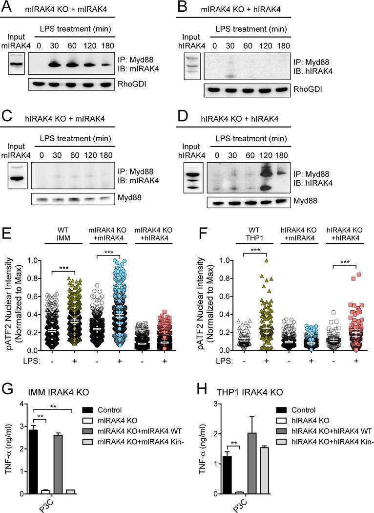
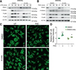
- Figure 6. IRAK4 kinase activity is dispensable for NF-kappaB and MAPK activation downstream of TLRs. A-C , BMDMs were pretreated with either DMSO or a 20 mu m concentration of a selective I4 inhb for 30 min. Cells were then left untreated or stimulated with 1 mug/ml LPS ( A ) or 500 ng/ml P3C ( B ) for an additional 30, 60, or 90 min. WCLs were then subjected to immunoblotting for IRAK1, P-IRAK4, NF-kappaB P-p65, P-p38 MAPK, and actin as a loading control. Data presented are representative of at least three independent experiments. C , cells were then left untreated ( UT ) or stimulated with 500 ng/ml P3C for an additional 4 h before fixation and staining with an NF-kappaB P-p65 antibody, followed by a specific secondary antibody conjugated to an Alexa-488 fluorophore. Samples were then examined by confocal microscopy. Representative images are presented from three biological replicates. Scale bar , 10 mum. D , NF-kappaB P-p65 nuclear translocation was then quantified by examining the mean fluorescence intensity of the Alexa-488 signal in cell nuclei. Nuclear intensity was made relative to DMSO-untreated samples and presented as the mean +- S.E. ( error bars ) (DMSO versus IRAK4 inhb: *, p < 0.05; **, p < 0.01) combined from BMDMs derived from three individual mice (biological replicates).