Explore
 Validate
 Learn
Learn Western blot
 Immunocytochemistry
Antibody data
- Antibody Data
- Antigen structure
- References [1]
- Comments [0]
- Validations
- Immunocytochemistry [7]
- Other assay [1]
Submit
Validation data
Reference
Comment
Report error
- Product number
- PA5-20018 - Provider product page

- Provider
- Invitrogen Antibodies
- Product name
- IRAK4 Polyclonal Antibody
- Antibody type
- Polyclonal
- Antigen
- Synthetic peptide
- Description
- In Western blot applications, this antibody detects a band at ~50kDa. A suggested positive control is K562 cell lysate. PA5-20018 can be used with blocking peptide PEP-0137.
- Reactivity
- Human
- Host
- Rabbit
- Isotype
- IgG
- Vial size
- 100 μg
- Concentration
- 1 mg/mL
- Storage
- Maintain refrigerated at 2-8°C for up to 3 months. For long term storage store at -20°C
Submitted references Fisetin Inhibits NLRP3 Inflammasome by Suppressing TLR4/MD2-Mediated Mitochondrial ROS Production.
Molagoda IMN, Athapaththu AMGK, Choi YH, Park C, Jin CY, Kang CH, Lee MH, Kim GY
Antioxidants (Basel, Switzerland) 2021 Jul 28;10(8)
Antioxidants (Basel, Switzerland) 2021 Jul 28;10(8)
No comments: Submit comment
Supportive validation
- Submitted by
- Invitrogen Antibodies (provider)
- Main image
- Experimental details
- Immunofluorescent analysis of K562 cells using a IRAK-4 polyclonal antibody (Product # PA5-20018) at a 10 µg/mL dilution.
- Submitted by
- Invitrogen Antibodies (provider)
- Main image

- Experimental details
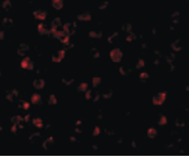
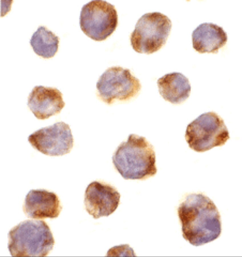
- Immunocytochemistry of K562 cells using IRAK4 Polyclonal Antibody (Product # PA5-20018) at 10 µg/mL. Cells were fixed with formaldehyde and blocked with 0.1 serum for 1 h at RT; antigen retrieval was by heat mediation with a citrate buffer (pH6). Samples were incubated with primary antibody overnight at 4°C. A goat anti-rabbit IgG H&L (HRP) at 1:250 was used as secondary. Counter stained with Hematoxylin.

- Submitted by
- Invitrogen Antibodies (provider)
- Main image
- Experimental details
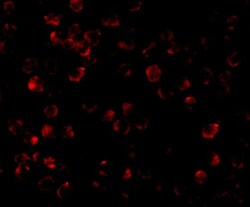
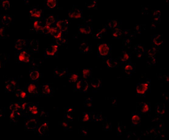
- Immunofluorescent analysis of 4% paraformaldehyde-fixed K562 Cells labeling IRAK4 with IRAK4 Polyclonal Antibody (Product # PA5-20018) at 10 µg/mL, followed by goat anti-rabbit IgG secondary antibody at 1:500 dilution (red).

- Submitted by
- Invitrogen Antibodies (provider)
- Main image

- Experimental details
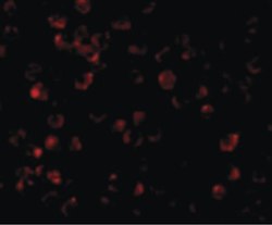
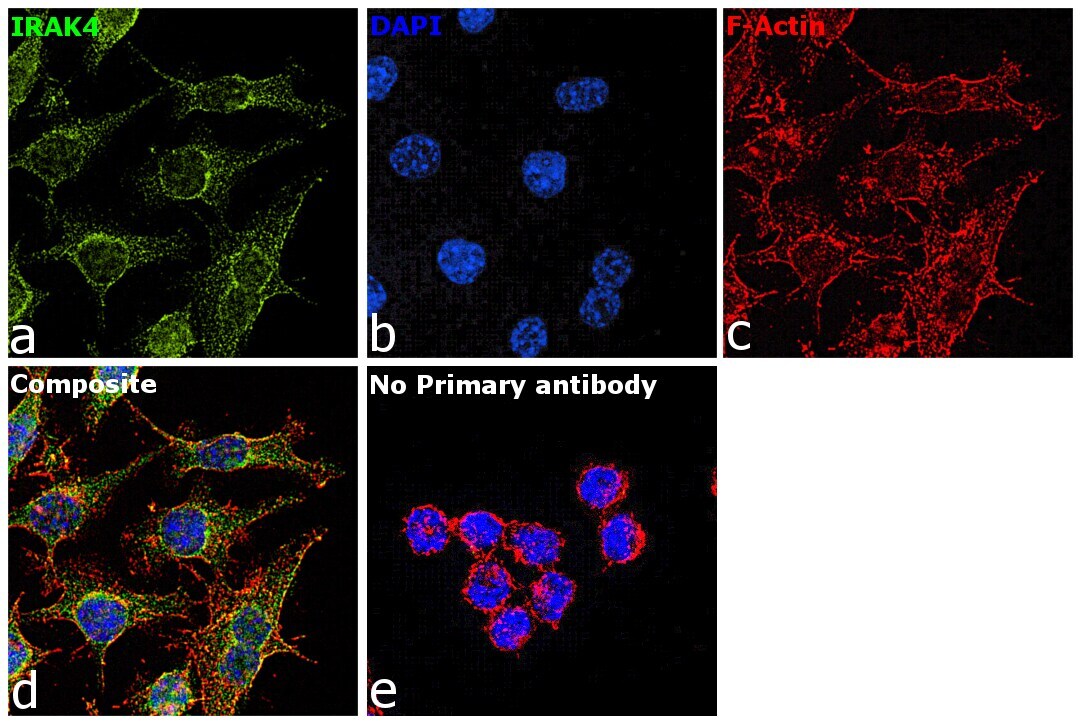
- Immunofluorescence analysis of IRAK4 was performed using RAW 264.7 cells. The cells were fixed with 4% paraformaldehyde for 10 minutes, permeabilized with 0.1% Triton™ X-100 for 15 minutes, and blocked with 2% BSA for 1 hour at room temperature. The cells were labeled with IRAK4 Polyclonal Antibody (Product # PA5-20018) at 5 µg/mL concentration in 0.1% BSA, incubated at 4 degree Celsius overnight and then with Goat anti-Rabbit IgG (H+L), Superclonal™ Recombinant Secondary Antibody, Alexa Fluor 488 conjugate (Product # A27034) at a dilution of 1:2000 for 45 minutes at room temperature (Panel a: green). Nuclei (Panel b: blue) were stained with SlowFade® Gold Antifade Mountant with DAPI (Product # S36938). F-actin (Panel c: red) was stained with Rhodamine Phalloidin (Product # R415, 1:300). Panel d represents the merged image showing staining in cytoplasm. Panel e represents control cells with no primary antibody to assess background. The images were captured at 60X magnification.

- Submitted by
- Invitrogen Antibodies (provider)
- Main image

- Experimental details
- Immunofluorescent analysis of 4% paraformaldehyde-fixed K562 Cells labeling IRAK4 with IRAK4 Polyclonal Antibody (Product # PA5-20018) at 10 µg/mL, followed by goat anti-rabbit IgG secondary antibody at 1:500 dilution (red).
- Submitted by
- Invitrogen Antibodies (provider)
- Main image

- Experimental details
- Immunocytochemistry of K562 cells using IRAK4 Polyclonal Antibody (Product # PA5-20018) at 10 µg/mL. Cells were fixed with formaldehyde and blocked with 0.1 serum for 1 h at RT; antigen retrieval was by heat mediation with a citrate buffer (pH6). Samples were incubated with primary antibody overnight at 4°C. A goat anti-rabbit IgG H&L (HRP) at 1:250 was used as secondary. Counter stained with Hematoxylin.
- Submitted by
- Invitrogen Antibodies (provider)
- Main image

- Experimental details
- Immunofluorescence analysis of IRAK4 was performed using RAW 264.7 cells. The cells were fixed with 4% paraformaldehyde for 10 minutes, permeabilized with 0.1% Triton™ X-100 for 15 minutes, and blocked with 2% BSA for 1 hour at room temperature. The cells were labeled with IRAK4 Polyclonal Antibody (Product # PA5-20018) at 5 µg/mL concentration in 0.1% BSA, incubated at 4 degree Celsius overnight and then with Goat anti-Rabbit IgG (Heavy Chain), Superclonal™ Recombinant Secondary Antibody, Alexa Fluor 488 conjugate (Product # A27034) at a dilution of 1:2000 for 45 minutes at room temperature (Panel a: green). Nuclei (Panel b: blue) were stained with SlowFade® Gold Antifade Mountant with DAPI (Product # S36938). F-actin (Panel c: red) was stained with Rhodamine Phalloidin (Product # R415, 1:300). Panel d represents the merged image showing staining in cytoplasm. Panel e represents control cells with no primary antibody to assess background. The images were captured at 60X magnification.
Supportive validation
- Submitted by
- Invitrogen Antibodies (provider)
- Main image
- Experimental details
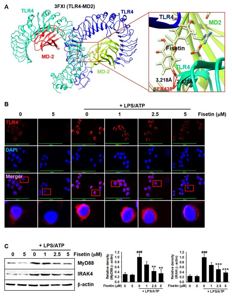
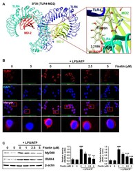
- Figure 2 Fisetin probably binds to TLR4 and inhibits the TLR4-mediated intracellular signaling pathway. ( A ) Molecular docking of fisetin (pose 1) with the TLR4/MD2 complex (PDB ID: 3FXI). ( B ) BV2 microglial cells were seeded on 3% gelatin-coated coverslips and treated with the indicated concentrations of fisetin (0-5 uM) for 2 h prior to stimulation with 1 ug/mL LPS for 2 h and subsequent stimulation with 1 mM ATP (LPS/ATP) for an additional 2 h. Cells were fixed with 4% paraformaldehyde and immunostained for TLR4 using Alexa Fluor 647-conjugated secondary antibody. The images were captured using a CELENA S Digital Imaging System. ( C ) In a parallel experiment, the total proteins were extracted and western blotting was performed for detecting the expression of MyD88 and IRAK4 proteins. beta-Actin was used as a loading control. The results indicate the mean +- standard error median (SEM), and is representative of the results obtained from three independent experiments. ### p < 0.001 vs. untreated cells; *** p < 0.001 and ** p < 0.01 vs. LPS/ATP-treated cells.