Explore
 Validate
 Learn
Learn Western blot
 Flow cytometry
Antibody data
- Antibody Data
- Antigen structure
- References [7]
- Comments [0]
- Validations
- Flow cytometry [3]
- Other assay [5]
Submit
Validation data
Reference
Comment
Report error
- Product number
- MA1-41280 - Provider product page

- Provider
- Invitrogen Antibodies
- Product name
- Caspase 8 Monoclonal Antibody (90A992)
- Antibody type
- Monoclonal
- Antigen
- Other
- Description
- MA1-41280 detects Caspase-8 in human, Rhesus monkey, and chimpanzee samples. Suggested positive control: Jurkat, antigen standard for CASP8 (transient overexpression lysate), Jurkat whole cell lysate.
- Reactivity
- Human, Drosophila
- Host
- Mouse
- Isotype
- IgG
- Antibody clone number
- 90A992
- Vial size
- 100 μg
- Concentration
- 1 mg/mL
- Storage
- Store at 4°C short term. For long term storage, store at -20°C, avoiding freeze/thaw cycles.
Submitted references Anti-Apoptotic Effect of Apelin in Human Placenta: Studies on BeWo Cells and Villous Explants from Third-Trimester Human Pregnancy.
Matrix Drug Screen Identifies Synergistic Drug Combinations to Augment SMAC Mimetic Activity in Ovarian Cancer.
In Vitro Effects of Vaspin on Porcine Granulosa Cell Proliferation, Cell Cycle Progression, and Apoptosis by Activation of GRP78 Receptor and Several Kinase Signaling Pathways Including MAP3/1, AKT, and STAT3.
Telomerase reverse transcriptase interference synergistically promotes tumor necrosis factor‑related apoptosis‑inducing ligand‑induced oral squamous cell carcinoma apoptosis and suppresses proliferation in vitro and in vivo.
5‑bromo‑3‑(3‑hydroxyprop‑1‑ynyl)‑2H‑pyran‑2‑one induces apoptosis in T24 human bladder cancer cells through mitochondria-dependent signaling pathways.
Esculetin induces antiproliferative and apoptotic response in pancreatic cancer cells by directly binding to KEAP1.
Dysregulation of the intrinsic apoptotic pathway mediates megakaryocytic hyperplasia in myeloproliferative neoplasms.
Mlyczyńska E, Myszka M, Kurowska P, Dawid M, Milewicz T, Bałajewicz-Nowak M, Kowalczyk P, Rak A
International journal of molecular sciences 2021 Mar 9;22(5)
International journal of molecular sciences 2021 Mar 9;22(5)
Matrix Drug Screen Identifies Synergistic Drug Combinations to Augment SMAC Mimetic Activity in Ovarian Cancer.
Noonan AM, Cousins A, Anderson D, Zeligs KP, Bunch K, Hernandez L, Shibuya Y, Goldlust IS, Guha R, Ferrer M, Thomas CJ, Annunziata CM
Cancers 2020 Dec 15;12(12)
Cancers 2020 Dec 15;12(12)
In Vitro Effects of Vaspin on Porcine Granulosa Cell Proliferation, Cell Cycle Progression, and Apoptosis by Activation of GRP78 Receptor and Several Kinase Signaling Pathways Including MAP3/1, AKT, and STAT3.
Kurowska P, Mlyczyńska E, Dawid M, Opydo-Chanek M, Dupont J, Rak A
International journal of molecular sciences 2019 Nov 19;20(22)
International journal of molecular sciences 2019 Nov 19;20(22)
Telomerase reverse transcriptase interference synergistically promotes tumor necrosis factor‑related apoptosis‑inducing ligand‑induced oral squamous cell carcinoma apoptosis and suppresses proliferation in vitro and in vivo.
Zhao X, Zhang C, Le Z, Zeng S, Pan C, Shi J, Wang J, Zhao X
International journal of molecular medicine 2018 Sep;42(3):1283-1294
International journal of molecular medicine 2018 Sep;42(3):1283-1294
5‑bromo‑3‑(3‑hydroxyprop‑1‑ynyl)‑2H‑pyran‑2‑one induces apoptosis in T24 human bladder cancer cells through mitochondria-dependent signaling pathways.
Yu GQ, Dou ZL, Jia ZH
Molecular medicine reports 2017 Jan;15(1):153-159
Molecular medicine reports 2017 Jan;15(1):153-159
Esculetin induces antiproliferative and apoptotic response in pancreatic cancer cells by directly binding to KEAP1.
Arora R, Sawney S, Saini V, Steffi C, Tiwari M, Saluja D
Molecular cancer 2016 Oct 18;15(1):64
Molecular cancer 2016 Oct 18;15(1):64
Dysregulation of the intrinsic apoptotic pathway mediates megakaryocytic hyperplasia in myeloproliferative neoplasms.
Malherbe JA, Fuller KA, Mirzai B, Kavanagh S, So CC, Ip HW, Guo BB, Forsyth C, Howman R, Erber WN
Journal of clinical pathology 2016 Apr 8;69(11):1017-24
Journal of clinical pathology 2016 Apr 8;69(11):1017-24
No comments: Submit comment
Supportive validation
- Submitted by
- Invitrogen Antibodies (provider)
- Main image
- Experimental details
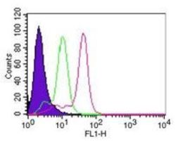
- Flow cytometric analysis of Caspase-8 in HeLa cells using 0.1 µg of Caspase 8 monoclonal antibody (Product # MA1-41280). Shaded histogram represents cells without antibody; green represents isotype control; red represents Caspase 8 monoclonal antibody (Product # MA1-41280). Goat anti-mouse IgG-FITC secondary antibody was used for this test.

- Submitted by
- Invitrogen Antibodies (provider)
- Main image

- Experimental details
- Flow cytometry of Caspase 8 in HeLa cells. Samples were incubated in Caspase 8 monoclonal antibody (Product # MA1-41280) using a dilution of 0.1 µg followed by Goat anti-mouse IgG-FITC secondary antibody. Shaded histogram represents cells without antibody; green represents isotype control; red represents Caspase-8 antibody. IC-Flow (Intracellular Staining Flow Cytometry Kit) was used to fix and prepare the cells for staining.

Supportive validation
- Submitted by
- Invitrogen Antibodies (provider)
- Main image
- Experimental details
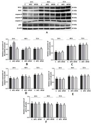
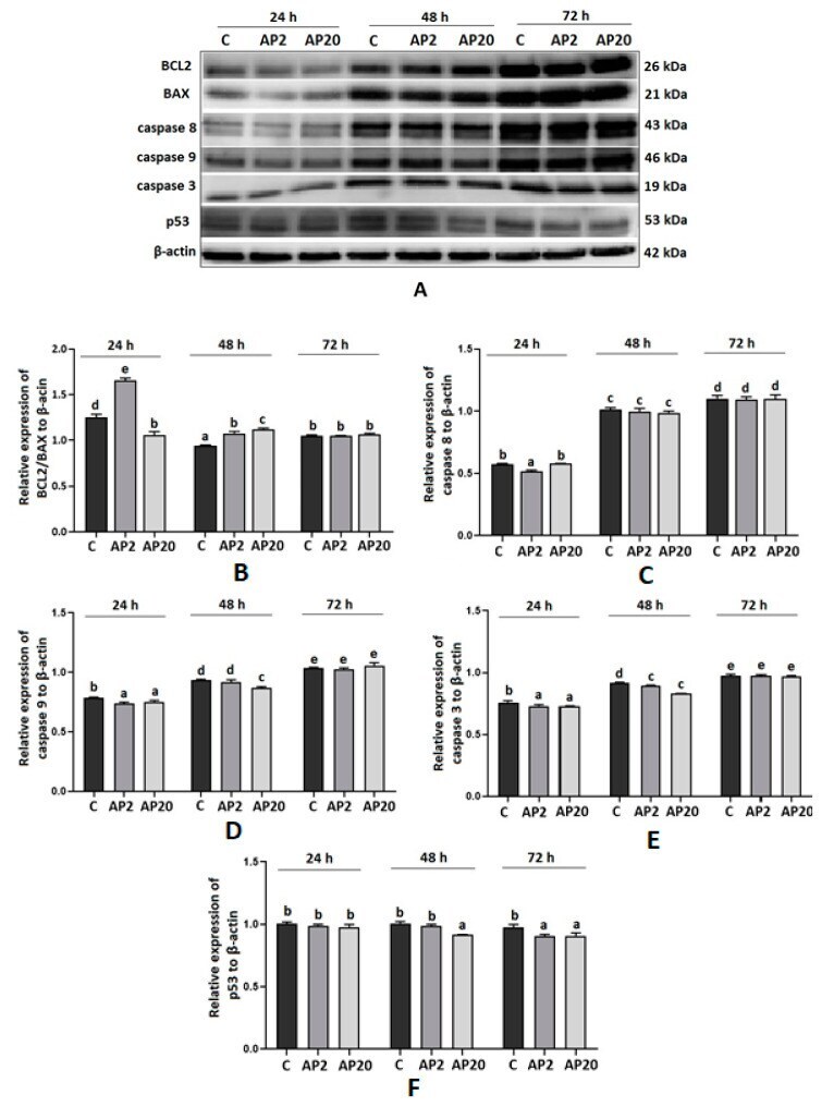
- Figure 1 Effect of apelin on protein expression of apoptotic factors in BeWo cells. The cells were incubated with apelin at doses 2 of (AP2) and 20 (AP20) ng/mL for 24, 48 and 72 h, and subsequently, Western blot analysis was performed to examine the expression of BCL2 (B-cell like lymphoma 2), BAX (Bcl-2-like protein 4), caspase 3, 8 and 9 and p53. Results are shown as stripes on gel image ( A ) and densitometry analysis relative to beta-actin ( B - F ). Experiments were independently performed and repeated three times ( n = 3). The data are arranged as means +- SEM. Different letters indicate significant differences ( p < 0.05) among groups; Control ( C ).
- Submitted by
- Invitrogen Antibodies (provider)
- Main image
- Experimental details
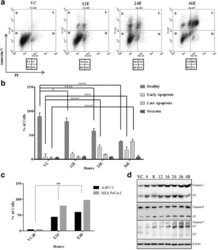
- Fig. 2 Esculetin induces apoptosis in pancreatic cancer cells: a Flow cytometric analysis of PANC-1 cells treated with 100 muM esculetin for indicated time showed temporal increase in surface expression of apoptotic marker- Annexin V indicating increased population of cells in apoptotic phase. b Percentage of PANC-1 cells exhibiting fluorescence in all four panels (healthy, early apoptosis, late apoptosis and necrosis) showing time dependent increase in apoptosis in Ecsuletin treated cells. c Percentage of cells with active APO BrdU indicating apoptosis in the absence and presence of esculetin (100 muM) as determined using TUNEL assay. d Western blot analysis showing an increase in expression of pro and active form of caspases (VC stands for vehicle control, E stands for esculetin treated sample for indicated time, CF stands for cleaved form, numerals represent time of esculetin treatment). Data represents the mean +- SD of three independent experiments. The significance was determined using ANOVA (Bonferroni's test). Key:* p < 0.05; ** p < 0.01; *** p < 0.001; **** p < 0.0001)

- Submitted by
- Invitrogen Antibodies (provider)
- Main image
- Experimental details
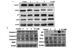
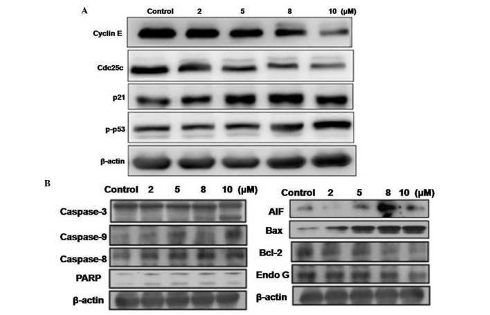
- Figure 5. BHP treatment alters expression levels of cell cycle proteins and apoptosis-inducing proteins in T24 cells. The cells were exposed to various concentrations of BHP and subjected to western blot analysis for (A) cell cycle and (B) apoptotic proteins. Cdc25c, cell division cycle 25C; p-p53, phosphorylated p53; PARP, poly(ADP-ribose) polymerase; AIF, apoptosis-inducing factor; Bcl-2, B cell lymphoma-2; Bax, Bcl-2-associated X protein; Endo G, endonuclease G.
- Submitted by
- Invitrogen Antibodies (provider)
- Main image
- Experimental details
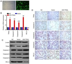
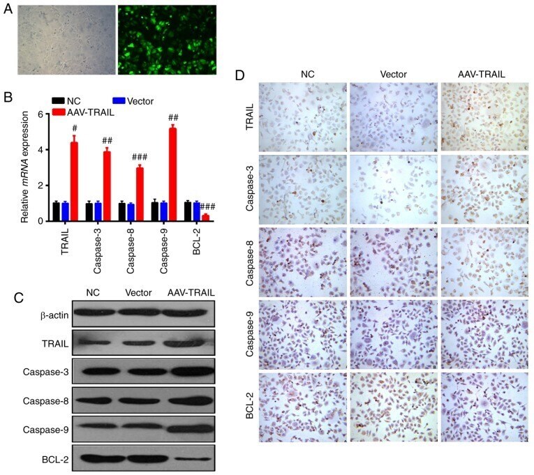
- Figure 1 AAV-mediated TRAIL overexpression induced increased expression of caspase-3, caspase-8, and caspase-9, and reduced expression of Bcl-2 in OSCC cells. (A) Immunofluorescence assay was carried out to detect the infection efficiency of the recombinant AAV system in the SCC25 OSCC cell line (magnification, x400). The left panel presents light microscopy and the right field presents fluorescence microscopy. (B) Reverse transcription-quantitative polymerase chain reaction, (C) western blotting and (D) immunocytochemistry analysis (magnification, x200) were performed to measure apoptosis-related genes expression changes in SCC25 cells with upregulation of TRAIL. Data are presented as the mean +- standard deviation. Each assay was performed independently at least three times. # P
- Submitted by
- Invitrogen Antibodies (provider)
- Main image
- Experimental details
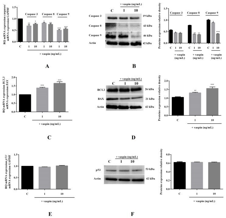
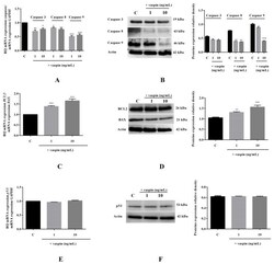
- Figure 5 Effect of vaspin on mRNA and protein expression levels of caspase-3, -8, and -9; BAX (bcl-2-like protein 4), and BCL2 (B-cell lymphoma 2). The cells were treated with 1 and 10 ng/mL vaspin for 24 h, and then, real-time PCR and Western blot analysis were performed to determine levels of caspase-3, -8, and -9 ( A , B ); BCL2 and BAX ( C , D ); and p53 ( E , F ) (full gel images are available in the Supplementary Materials ). Gene expression levels were normalised to glyceraldehyde 3-phosphate dehydrogenase ( GAPDH) , while proteins levels were normalized to actin. Experiments were independently performed and repeated five times ( n = 5). The data are plotted as the means +- SEM. Significance between control and vaspin treatments is indicated by * p < 0.05, ** p < 0.01 and *** p < 0.001; Control (C).