 Explore
 Validate
 Learn
Learn Western blot
 ELISA
ELISA Other assay
Antibody data
- Antibody Data
- Antigen structure
- References [1]
- Comments [0]
- Validations
- Other assay [1]
Submit
Validation data
Reference
Comment
Report error
- Product number
- MA1-20168 - Provider product page

- Provider
- Invitrogen Antibodies
- Product name
- FADD Monoclonal Antibody (FD19)
- Antibody type
- Monoclonal
- Antigen
- Purifed from natural sources
- Description
- Recommended positive controls: A431. Store product as a concentrated solution. Centrifuge briefly prior to opening the vial.
- Reactivity
- Human, Rat
- Host
- Mouse
- Isotype
- IgG
- Antibody clone number
- FD19
- Vial size
- 100 μL
- Concentration
- 2.1 mg/mL
- Storage
- Store at 4°C short term. For long term storage, store at -20°C, avoiding freeze/thaw cycles.
Submitted references Induction of interferon-β and interferon signaling by TRAIL and Smac mimetics via caspase-8 in breast cancer cells.
Granqvist V, Holmgren C, Larsson C
PloS one 2021;16(3):e0248175
PloS one 2021;16(3):e0248175
No comments: Submit comment
Supportive validation
- Submitted by
- Invitrogen Antibodies (provider)
- Main image
- Experimental details
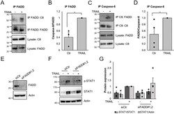
- Fig 6 Involvement of FADD in TRAIL-mediated IFN signaling. A-D) MCF-7 cells were stimulated with 100 ng/ml TRAIL for 2 h in the presence of 20 muM zVAD-FMK and lysates were immunoprecipitated with antibodies towards FADD (A, B) or caspase-8 (C, D). Lysates and immunoprecipitates were subjected to Western blot for FADD and caspase-8 (A and C). Intensities in bands for the immunoprecipitates were quantified (B and D). E-G) MCF-7 cells were transfected for 72 h with a combination of two siRNAs targeting FADD (E) followed by treatment with TRAIL (100 ng/ml) for 48h. Lysates were subjected to Western blot with indicated antibodies (F) and intensities in bands were quantified (G). Data (B,D,G) are mean +- SEM with individual data points indicated and experiments were performed three (B) or four (D,G) times. * denotes p
