Explore
 Validate
 Learn
Learn Western blot
 Immunohistochemistry
Antibody data
- Antibody Data
- Antigen structure
- References [1]
- Comments [0]
- Validations
- Western blot [1]
Submit
Validation data
Reference
Comment
Report error
- Product number
- A00237-3 - Provider product page

- Provider
- Boster Biological Technology
- Product name
- Anti-FADD Antibody Picoband™
- Antibody type
- Polyclonal
- Description
- Rabbit IgG polyclonal antibody for FADD detection. Tested with WB, IHC-P, FCM in Mouse;Rat.
- Reactivity
- Mouse, Rat
- Host
- Rabbit
- Vial size
- 100μg/vial
- Concentration
- Add 0.2ml of distilled water will yield a concentration of 500μg/ml.
- Storage
- At -20°C for one year. After reconstitution, at 4°C for one month. It can also be aliquoted and stored frozen at -20°C for a longer time. Avoid repeated freezing and thawing.
- Handling
- Add 0.2ml of distilled water will yield a concentration of 500μg/ml.
Submitted references Aconitine induces cell apoptosis via mitochondria and death receptor signaling pathways in hippocampus cell line.
Wang H, Liu Y, Guo Z, Wu K, Zhang Y, Tian Y, Zhao B, Lu H
Research in veterinary science 2022 Mar;143:124-133
Research in veterinary science 2022 Mar;143:124-133
No comments: Submit comment
Supportive validation
- Submitted by
- Boster Biological Technology (provider)
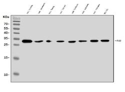
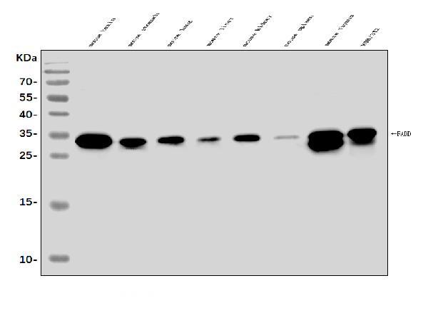
- Main image
- Experimental details
- Western blot analysis of FADD using anti-FADD antibody (A00237-3). Electrophoresis was performed on a 5-20% SDS-PAGE gel at 70V (Stacking gel) / 90V (Resolving gel) for 2-3 hours. The sample well of each lane was loaded with 30ug of sample under reducing conditions. Lane 1: mouse brain tissue lysates, Lane 2: mouse stomach tissue lysates, Lane 3: mouse lung tissue lysates, Lane 4: mouse liver tissue lysates, Lane 5: mouse kidney tissue lysates, Lane 6: mouse spleen tissue lysates, Lane 7: mouse thymus tissue lysates, Lane 8: mouse NIH/3T3 whole cell lysates. After Electrophoresis, proteins were transferred to a Nitrocellulose membrane at 150mA for 50-90 minutes. Blocked the membrane with 5% Non-fat Milk/ TBS for 1.5 hour at RT. The membrane was incubated with rabbit anti-FADD antigen affinity purified polyclonal antibody (Catalog # A00237-3) at 0.25 μg/mL overnight at 4°C, then washed with TBS-0.1%Tween 3 times with 5 minutes each and probed with a goat anti-rabbit IgG-HRP secondary antibody at a dilution of 1:5000 for 1.5 hour at RT. The signal is developed using an Enhanced Chemiluminescent detection (ECL) kit (Catalog # EK1002) with Tanon 5200 system. A specific band was detected for FADD at approximately 30KD. The expected band size for FADD is at 23KD.
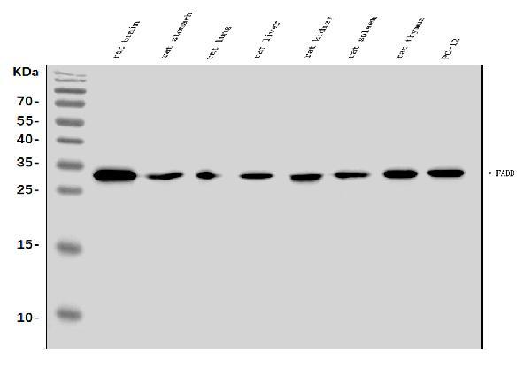
- Additional image