 Explore
 Validate
 Learn
Learn Western blot
 ELISA
ELISAAntibody data
- Antibody Data
- Antigen structure
- References [1]
- Comments [0]
- Validations
- Western blot [1]
- Immunocytochemistry [2]
- Immunohistochemistry [1]
- Flow cytometry [1]
Submit
Validation data
Reference
Comment
Report error
- Product number
- NB300-996 - Provider product page

- Provider
- Novus Biologicals
- Proper citation
- Novus Cat#NB300-996, RRID:AB_2257356
- Product name
- Goat Polyclonal WASP Antibody
- Antibody type
- Polyclonal
- Description
- Immunogen affinity purified. No cross-reactivity expected with N WASP (WASL).
- Reactivity
- Human
- Host
- Goat
- Isotype
- IgG
- Vial size
- 0.1 mg
- Concentration
- 0.5 mg/ml
- Storage
- Store at -20C. Avoid freeze-thaw cycles.
Submitted references Molecular mechanisms of invadopodium formation: the role of the N-WASP-Arp2/3 complex pathway and cofilin.
Yamaguchi H, Lorenz M, Kempiak S, Sarmiento C, Coniglio S, Symons M, Segall J, Eddy R, Miki H, Takenawa T, Condeelis J
The Journal of cell biology 2005 Jan 31;168(3):441-52
The Journal of cell biology 2005 Jan 31;168(3):441-52
No comments: Submit comment
Supportive validation
- Submitted by
- Novus Biologicals (provider)
- Main image

- Experimental details
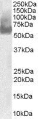
- Western Blot: WASP Antibody [NB300-996] - U937 lysate (RIPA buffer, 30 ug total protein). Antibody at 0.03 ug/mL. Detected by chemiluminescence.
Supportive validation
- Submitted by
- Novus Biologicals (provider)
- Main image
- Experimental details
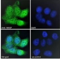
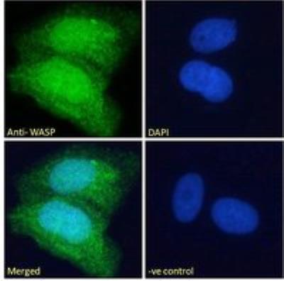
- Immunocytochemistry/Immunofluorescence
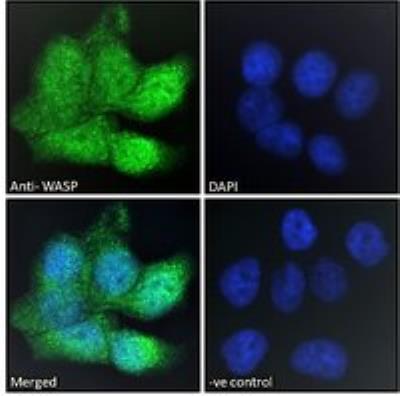
- Submitted by
- Novus Biologicals (provider)
- Main image
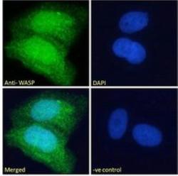
- Experimental details
- Immunocytochemistry/Immunofluorescence
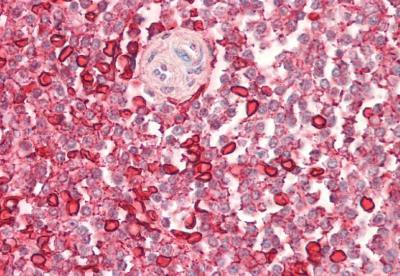
Supportive validation
- Submitted by
- Novus Biologicals (provider)
- Main image
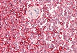
- Experimental details
- Immunohistochemistry-Paraffin: WASP Antibody [NB300-996] - Staining of paraffin embedded Human Spleen. Antibody at 5 ug/mL. Steamed antigen retrieval with citrate buffer pH 6, AP-staining.
Supportive validation
- Submitted by
- Novus Biologicals (provider)
- Main image
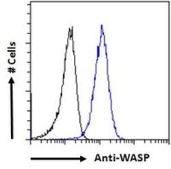
- Experimental details
- Flow Cytometry: WASP Antibody [NB300-996] - Flow cytometric analysis of paraformaldehyde fixed HepG2 cells (blue line), permeabilized with 0.5% Triton. Primary incubation 1hr (10 ug/mL) followed by Alexa Fluor 488 secondary antibody (1 ug/mL). IgG control: Unimmunized goat IgG (black line) followed by Alexa Fluor 488 secondary antibody.
