Explore
 Validate
 Learn
Learn Western blot
Antibody data
- Antibody Data
- Antigen structure
- References [0]
- Comments [0]
- Validations
- Western blot [2]
- Immunocytochemistry [3]
Submit
Validation data
Reference
Comment
Report error
- Product number
- PA5-21532 - Provider product page

- Provider
- Invitrogen Antibodies
- Product name
- CD55 Polyclonal Antibody
- Antibody type
- Polyclonal
- Antigen
- Recombinant full-length protein
- Description
- Recommended positive controls: K562, THP-1. Store product as a concentrated solution. Centrifuge briefly prior to opening the vial.
- Reactivity
- Human
- Host
- Rabbit
- Isotype
- IgG
- Vial size
- 100 μL
- Concentration
- 1 mg/mL
- Storage
- Store at 4°C short term. For long term storage, store at -20°C, avoiding freeze/thaw cycles.
No comments: Submit comment
Supportive validation
- Submitted by
- Invitrogen Antibodies (provider)
- Main image

- Experimental details
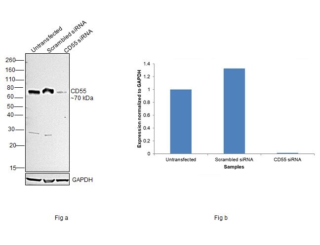
- Knockdown of Complement decay-accelerating factor was achieved by transfecting HeLa with Complement decay-accelerating factor specific siRNAs (Silencer® select Product # S534833, S3906). Western Blot analysis (Fig. a) was performed using Whole cell extracts from the Complement decay-accelerating factor knockdown cells (lane 3), non-targeting scrambled siRNA transfected cells (lane 2) and untransfected cells (lane 1). The Blot was probed with CD55 Polyclonal Antibody (Product # PA5-21532, 1:1000 dilution) and Goat anti-Rabbit IgG (Heavy Chain) Superclonal™ Recombinant Secondary Antibody, HRP (Product # A27036, 1:4000 dilution). Densitometric analysis of this western Blot is shown in histogram (Fig. b). Decrease in signal upon siRNA mediated knock down confirms that antibody is specific to Complement decay-accelerating factor.
- Submitted by
- Invitrogen Antibodies (provider)
- Main image

- Experimental details
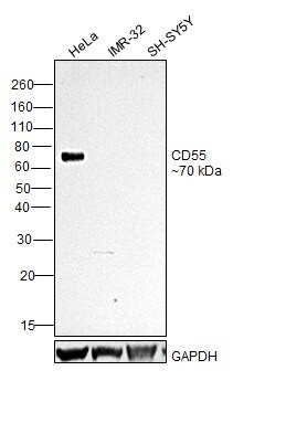
- Western Blot was performed using Anti-CD55 Polyclonal Antibody (Product # PA5-21532) and a 70 kDa band corresponding to Complement decay-accelerating factor was observed across positive cell line (HeLa); and not across negative cell lines (IMR-32 and SH-SY5Y). Whole cell extracts (30 µg lysate) of HeLa (Lane 1), IMR-32 (Lane 2) and SH-SY5Y (Lane 3) were electrophoresed using NuPAGE™ 10% Bis-Tris Protein Gel (Product # NP0302BOX). Resolved proteins were then transferred onto a Nitrocellulose membrane (Product # IB23001) by iBlot® 2 Dry Blotting System (Product # IB21001). The Blot was probed with the primary antibody (1:1000 dilution) and detected by chemiluminescence with Goat anti-Rabbit IgG (Heavy Chain) Superclonal™ Recombinant Secondary Antibody, HRP (Product # A27036, 1:4000 dilution) using the iBright FL 1000 (Product # A32752). Chemiluminescent detection was performed using Novex® ECL Chemiluminescent Substrate Reagent Kit (Product # WP20005).
Supportive validation
- Submitted by
- Invitrogen Antibodies (provider)
- Main image

- Experimental details
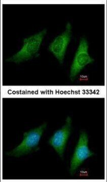
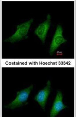
- Immunofluorescent analysis of CD55/DAF in methanol-fixed HeLa cells using a CD55/DAF polyclonal antibody (Product # PA5-21532) at a 1:200 dilution.
- Submitted by
- Invitrogen Antibodies (provider)
- Main image

- Experimental details
- Immunofluorescence analysis of methanol-fixed HeLa, using CD55 antibody (Product # PA5-21532) at 1:200 dilution.

- Submitted by
- Invitrogen Antibodies (provider)
- Main image

- Experimental details
- Immunofluorescence analysis of methanol-fixed HeLa, using CD55 antibody (Product # PA5-21532) at 1:200 dilution.