Explore
 Validate
 Learn
Learn Western blot
 Immunocytochemistry
Antibody data
- Antibody Data
- Antigen structure
- References [2]
- Comments [0]
- Validations
- Immunocytochemistry [1]
Submit
Validation data
Reference
Comment
Report error
- Product number
- BAF2009 - Provider product page

- Provider
- R&D Systems
- Product name
- Human CD55/DAF Biotinylated Antibody
- Antibody type
- Polyclonal
- Description
- Antigen Affinity-purified. Detects CD55/DAF in Western blots.
- Reactivity
- Human
- Host
- Goat
- Conjugate
- Biotin
- Antigen sequence
P08174- Isotype
- IgG
- Vial size
- 50 ug
- Concentration
- LYOPH
- Storage
- Use a manual defrost freezer and avoid repeated freeze-thaw cycles. 12 months from date of receipt, -20 to -70 °C as supplied. 1 month, 2 to 8 °C under sterile conditions after reconstitution. 6 months, -20 to -70 °C under sterile conditions after reconstitution.
Submitted references Complement inhibition ameliorates blast-induced acute lung injury in rats: Potential role of complement in intracellular HMGB1-mediated inflammation.
Decay-accelerating factor attenuates remote ischemia-reperfusion-initiated organ damage.
Li Y, Yang Z, Chavko M, Liu B, Aderemi OA, Simovic MO, Dubick MA, Cancio LC
PloS one 2018;13(8):e0202594
PloS one 2018;13(8):e0202594
Decay-accelerating factor attenuates remote ischemia-reperfusion-initiated organ damage.
Weeks C, Moratz C, Zacharia A, Stracener C, Egan R, Peckham R, Moore FD Jr, Tsokos GC
Clinical immunology (Orlando, Fla.) 2007 Sep;124(3):311-27
Clinical immunology (Orlando, Fla.) 2007 Sep;124(3):311-27
No comments: Submit comment
Supportive validation
- Submitted by
- R&D Systems (provider)
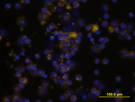
- Main image
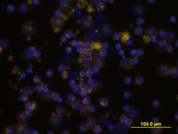
- Experimental details
- CD55/DAF in Human PBMCs. CD55/DAF was detected in immersion fixed human peripheral blood mononuclear cells (PBMCs) using Goat Anti-Human CD55/DAF Biotinylated Antigen Affinity-purified Polyclonal Antibody (Catalog # BAF2009) at 10 µg/mL for 3 hours at room temperature. Cells were stained using the NorthernLights™ 557-conjugated Streptavidin (yellow; Catalog # NL999) and counterstained with DAPI (blue). View our protocol for Fluorescent ICC Staining of Non-adherent Cells.