 Explore
 Validate
 Learn
LearnPA5-19701
antibody from Invitrogen Antibodies
Targeting: SNAP25
bA416N4.2, dJ1068F16.2, RIC-4, RIC4, SEC9, SNAP, SNAP-25
 Western blot
 Immunocytochemistry
 Immunoprecipitation
Antibody data
- Antibody Data
- Antigen structure
- References [0]
- Comments [0]
- Validations
- Immunocytochemistry [5]
Submit
Validation data
Reference
Comment
Report error
- Product number
- PA5-19701 - Provider product page

- Provider
- Invitrogen Antibodies
- Product name
- SNAP25 Polyclonal Antibody
- Antibody type
- Polyclonal
- Antigen
- Synthetic peptide
- Description
- This antibody is predicted to react with chicken and cow based on sequence homology. Store antibody at 4ºC for 1-2 weeks. For long-term storage, store at -20ºC.
- Reactivity
- Human, Mouse, Rat, Zebrafish
- Host
- Rabbit
- Isotype
- IgG
- Vial size
- 100 μg
- Concentration
- 1 mg/mL
- Storage
- -20°C or -80°C if preferred
No comments: Submit comment
Supportive validation
- Submitted by
- Invitrogen Antibodies (provider)
- Main image
- Experimental details
- Immunofluorescent staining of SH-SY5Y cells using Product # PA5-19701, anti-SNAP25 antibody. The cells were fixed with PFA (4%) for 10 minutes, permabilised with BSA (1%), normal goat serum (10%) and glycine (0.3 M) in 0.1% T-BST for 20 minutes and exposed to the primary antibody at a concentration of 5 µg/mL for 1 hour at room temp. The secondary antibody was a 448 fluorescence conjugated Goat anti-rabbit IgG (green) at a dilution of 1:1000. A WGA- 594 fluorescent conjugated stain was used to label plasma membranes (red) and the nuclei stain was DAPI (blue).

- Submitted by
- Invitrogen Antibodies (provider)
- Main image
- Experimental details
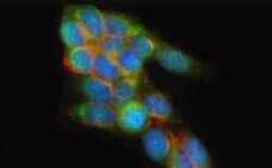
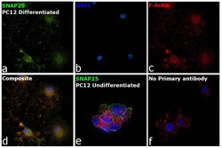
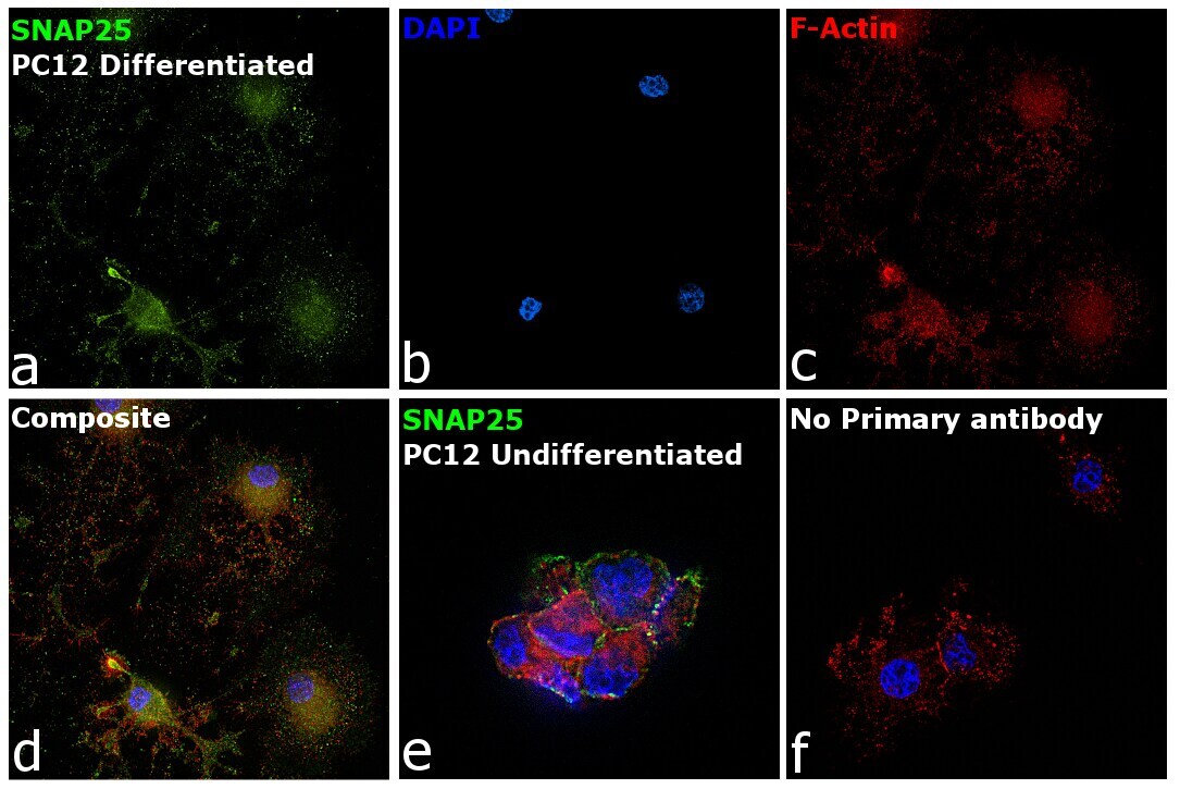
- Immunofluorescence analysis of Synaptosomal-associated protein 25 was performed using 70% confluent log phase PC-12 cells. The cells were fixed with 4% paraformaldehyde for 10 minutes, permeabilized with 0.1% Triton™ X-100 for 15 minutes, and blocked with 2% BSA for 45 minutes at room temperature. The cells were labeled with SNAP25 Polyclonal Antibody (Product # PA5-19701) at 0.8 µg/mL in 0.1% BSA, incubated at 4 degree celsius overnight and then labeled with Goat anti-Rabbit IgG (H+L) Superclonal™ Recombinant Secondary Antibody, Alexa Fluor® 488 conjugate (Product # A27034), (1:2000 dilution), for 45 minutes at room temperature (Panel a: Green). Nuclei (Panel b: Blue) were stained with ProLong™ Diamond Antifade Mountant with DAPI (Product # P36962). F-actin (Panel c: Red) was stained with Rhodamine Phalloidin (Product # R415, 1:300). Panel d represents the merged image showing enhanced cytoplasmic and membrane localization of SNAP-25 in PC-12 cells differentiated to neuronal phenotype with NGF (200 ng/mL 5 days). Panel e represents the undifferentiated PC-12 cells. Panel f represents control cells with no primary antibody to assess background. The images were captured at 60X magnification.
- Submitted by
- Invitrogen Antibodies (provider)
- Main image

- Experimental details
- Immunofluorescent staining of SH-SY5Y cells using Product # PA5-19701, anti-SNAP25 antibody. The cells were fixed with PFA (4%) for 10 minutes, permabilised with BSA (1%), normal goat serum (10%) and glycine (0.3 M) in 0.1% T-BST for 20 minutes and exposed to the primary antibody at a concentration of 5 µg/mL for 1 hour at room temp. The secondary antibody was a 448 fluorescence conjugated Goat anti-rabbit IgG (green) at a dilution of 1:1000. A WGA- 594 fluorescent conjugated stain was used to label plasma membranes (red) and the nuclei stain was DAPI (blue).

- Submitted by
- Invitrogen Antibodies (provider)
- Main image

- Experimental details
- Immunofluorescence analysis of Synaptosomal-associated protein 25 was performed using 70% confluent log phase PC-12 cells. The cells were fixed with 4% paraformaldehyde for 10 minutes, permeabilized with 0.1% Triton™ X-100 for 15 minutes, and blocked with 2% BSA for 45 minutes at room temperature. The cells were labeled with SNAP25 Polyclonal Antibody (Product # PA5-19701) at 0.8 µg/mL in 0.1% BSA, incubated at 4 degree celsius overnight and then labeled with Goat anti-Rabbit IgG (Heavy Chain) Superclonal™ Recombinant Secondary Antibody, Alexa Fluor® 488 conjugate (Product # A27034), (1:2000 dilution), for 45 minutes at room temperature (Panel a: Green). Nuclei (Panel b: Blue) were stained with ProLong™ Diamond Antifade Mountant with DAPI (Product # P36962). F-actin (Panel c: Red) was stained with Rhodamine Phalloidin (Product # R415, 1:300). Panel d represents the merged image showing enhanced cytoplasmic and membrane localization of SNAP-25 in PC-12 cells differentiated to neuronal phenotype with NGF (200 ng/mL 5 days). Panel e represents the undifferentiated PC-12 cells. Panel f represents control cells with no primary antibody to assess background. The images were captured at 60X magnification.
