Explore
 Validate
 Learn
Learn Western blot
 Immunocytochemistry
Antibody data
- Antibody Data
- Antigen structure
- References [4]
- Comments [0]
- Validations
- Western blot [2]
- Immunohistochemistry [3]
Submit
Validation data
Reference
Comment
Report error
- Product number
- NBP2-15039 - Provider product page

- Provider
- Novus Biologicals
- Product name
- Rabbit Polyclonal NFIX Antibody
- Antibody type
- Polyclonal
- Description
- Immunogen affinity purified.
- Reactivity
- Human, Mouse, Rat
- Host
- Rabbit
- Isotype
- IgG
- Vial size
- 0.1 ml
- Storage
- Aliquot and store at -20C or -80C. Avoid freeze-thaw cycles.
Submitted references Nuclear factor IX promotes glioblastoma development through transcriptional activation of Ezrin.
MiR-663a Stimulates Proliferation and Suppresses Early Apoptosis of Human Spermatogonial Stem Cells by Targeting NFIX and Regulating Cell Cycle.
RhoA and ERK signalling regulate the expression of the transcription factor Nfix in myogenic cells.
Nfix Induces a Switch in Sox6 Transcriptional Activity to Regulate MyHC-I Expression in Fetal Muscle.
Liu Z, Ge R, Zhou J, Yang X, Cheng KK, Tao J, Wu D, Mao J
Oncogenesis 2020 Apr 14;9(4):39
Oncogenesis 2020 Apr 14;9(4):39
MiR-663a Stimulates Proliferation and Suppresses Early Apoptosis of Human Spermatogonial Stem Cells by Targeting NFIX and Regulating Cell Cycle.
Zhou F, Yuan Q, Zhang W, Niu M, Fu H, Qiu Q, Mao G, Wang H, Wen L, Wang H, Lu M, Li Z, He Z
Molecular therapy. Nucleic acids 2018 Sep 7;12:319-336
Molecular therapy. Nucleic acids 2018 Sep 7;12:319-336
RhoA and ERK signalling regulate the expression of the transcription factor Nfix in myogenic cells.
Taglietti V, Angelini G, Mura G, Bonfanti C, Caruso E, Monteverde S, Le Carrou G, Tajbakhsh S, Relaix F, Messina G
Development (Cambridge, England) 2018 Oct 29;145(21)
Development (Cambridge, England) 2018 Oct 29;145(21)
Nfix Induces a Switch in Sox6 Transcriptional Activity to Regulate MyHC-I Expression in Fetal Muscle.
Taglietti V, Maroli G, Cermenati S, Monteverde S, Ferrante A, Rossi G, Cossu G, Beltrame M, Messina G
Cell reports 2016 Nov 22;17(9):2354-2366
Cell reports 2016 Nov 22;17(9):2354-2366
No comments: Submit comment
Supportive validation
- Submitted by
- Novus Biologicals (provider)
- Main image
- Experimental details
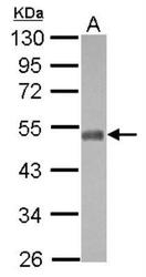
- Western Blot: NFIX Antibody [NBP2-15039] - Sample (30 ug of whole cell lysate) A: U87-MG 10% SDS PAGE diluted at 1:1000 The HRP-conjugated anti-rabbit IgG antibody (NBP2-19301 was used to detect the primary antibody.

- Submitted by
- Novus Biologicals (provider)
- Main image
- Experimental details
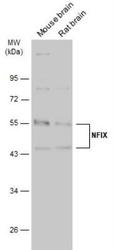
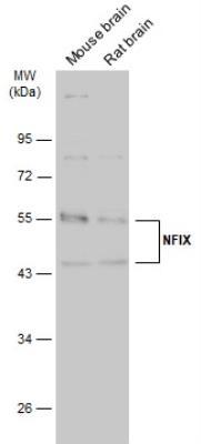
- Western Blot: NFIX Antibody [NBP2-15039] - Various tissue extracts (50 ug) were separated by 10% SDS-PAGE, and the membrane was blotted with NFIX antibody [N3C3] diluted at 1:1000. The HRP-conjugated anti-rabbit IgG antibody (NBP2-19301) was used to detect the primary antibody.
Supportive validation
- Submitted by
- Novus Biologicals (provider)
- Main image
- Experimental details
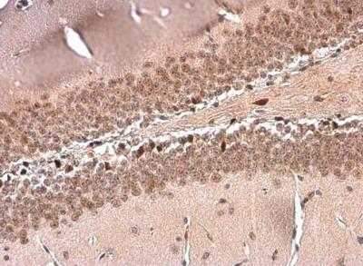
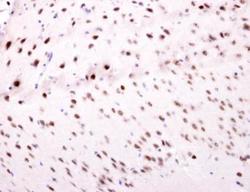
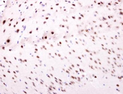
- Immunohistochemistry-Paraffin: NFIX Antibody [NBP2-15039] - Paraffin-embedded mouse fore brain. NFIX antibody [N3C3] dilution: 1:500.
- Submitted by
- Novus Biologicals (provider)
- Main image
- Experimental details
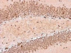
- Immunohistochemistry-Paraffin: NFIX Antibody [NBP2-15039] - Mouse hippocampus. NFIX stained by NFIX antibody [N3C3] diluted at 1:500.
- Submitted by
- Novus Biologicals (provider)
- Main image
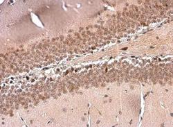
- Experimental details
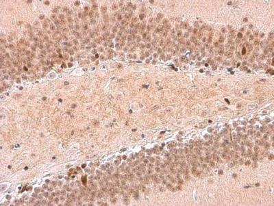
- Immunohistochemistry-Paraffin: NFIX Antibody [NBP2-15039] - Mouse hippocampus. NFIX stained by NFIX antibody [N3C3] diluted at 1:500.