Explore
 Validate
 Learn
Learn Western blot
 Immunocytochemistry
Antibody data
- Antibody Data
- Antigen structure
- References [1]
- Comments [0]
- Validations
- Immunocytochemistry [2]
- Other assay [2]
Submit
Validation data
Reference
Comment
Report error
- Product number
- 720124 - Provider product page

- Provider
- Invitrogen Antibodies
- Product name
- TYK2 Polyclonal Antibody
- Antibody type
- Polyclonal
- Antigen
- Recombinant full-length protein
- Description
- These Polyclonal antibodies are of rabbit origin developed by immunizing animals with proteins or peptides. The polyclonal antibody is purified by affinity purification from the rabbit sera generated after immunizing the rabbits with a specific type of protein or peptide. The purified antibody is tested for its functionality in various relevant research applications. The antibody is developed for Research Use Only and is non-hazardous or non-infectious in nature.
- Reactivity
- Human
- Host
- Rabbit
- Isotype
- IgG
- Vial size
- 100 μg
- Concentration
- 0.5 mg/mL
- Storage
- Store at 4°C short term. For long term storage, store at -20°C, avoiding freeze/thaw cycles.
Submitted references TYK2 promotes malignant peripheral nerve sheath tumor progression through inhibition of cell death.
Qin W, Godec A, Zhang X, Zhu C, Shao J, Tao Y, Bu X, Hirbe AC
Cancer medicine 2019 Sep;8(11):5232-5241
Cancer medicine 2019 Sep;8(11):5232-5241
No comments: Submit comment
Supportive validation
- Submitted by
- Invitrogen Antibodies (provider)
- Main image
- Experimental details
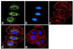
- Immunofluorescence was performed on fixed and permeabilized A549 cells for detection of TYK-2 using Anti-TYK-2 Rabbit Polyclonal Antibody (Product # 720124, 1 µg/mL) and labeled with Goat anti-Rabbit IgG (H+L) Superclonal Secondary Antibody, Alexa Fluor® 488 conjugate (Product # A27034, 1:2000). Panel a) shows representative cells that were stained for detection and localization of TYK2 protein (green), Panel b) is stained for nuclei (blue) using SlowFade® Gold Antifade Mountant with DAPI (Product # S36938). Panel c) represents cytoskeletal F-actin staining using Alexa Fluor® 555 Rhodamine Phalloidin (Product # R415, 1:300). Panel d) is a composite image of Panels a, b and c clearly demonstrating Cytoplasmic localization of TYK-2. Panel e) represents control cells with no primary Antibody to assess background.
- Submitted by
- Invitrogen Antibodies (provider)
- Main image

- Experimental details
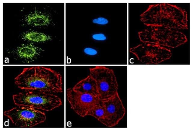
- Immunofluorescence was performed on fixed and permeabilized A549 cells for detection of TYK-2 using Anti-TYK-2 Rabbit Polyclonal Antibody (Product # 720124, 1 µg/mL) and labeled with Goat anti-Rabbit IgG (Heavy Chain) Superclonal Secondary Antibody, Alexa Fluor® 488 conjugate (Product # A27034, 1:2000). Panel a) shows representative cells that were stained for detection and localization of TYK2 protein (green), Panel b) is stained for nuclei (blue) using SlowFade® Gold Antifade Mountant with DAPI (Product # S36938). Panel c) represents cytoskeletal F-actin staining using Alexa Fluor® 555 Rhodamine Phalloidin (Product # R415, 1:300). Panel d) is a composite image of Panels a, b and c clearly demonstrating Cytoplasmic localization of TYK-2. Panel e) represents control cells with no primary Antibody to assess background.

Supportive validation
- Submitted by
- Invitrogen Antibodies (provider)
- Main image
- Experimental details
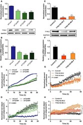
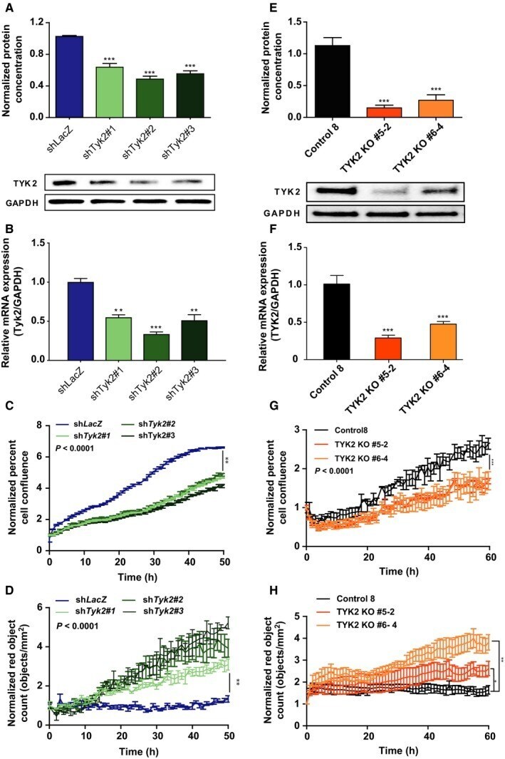
- Figure 2 Loss of Tyk2 /TYK2 in JW23.3 murine MPNST and MPNST 724 cells leads to increased cell death. sh Tyk2 -infected JW23.3 cell lines and control sh LacZ : (A) Western blot densitometry analysis measuring TYK2 protein. (B) Relative Expression of Tyk2 mRNA compared to Gapdh . (C) An Incucyte cell proliferation assay measuring confluence over time. (D) An Incucyte death assay measuring TOTO TM -3 iodide fluorescence as an indicator of death over time. TYK2 knockout MPNST 724 cells and scramble control: (E) Western blot densitometry analysis of TYK2 protein. (F) Relative Expression of TYK2 mRNA compared to GAPDH. (G) An Incucyte cell proliferation assay measuring confluence over time. (H) An Incucyte death assay measuring TOTO TM -3 iodide fluorescence as an indicator of death over time. (* P < 0.05, ** P < 0.01, *** P < 0.001)
- Submitted by
- Invitrogen Antibodies (provider)
- Main image

- Experimental details
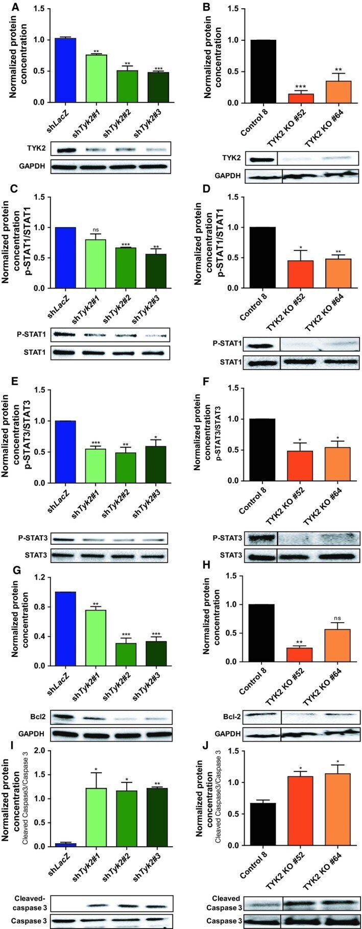
- Figure 3 Knockdown of Tyk2 / TYK2 in JW23.3 murine MPNST and MPNST 724 cells affects expression of downstream targets. Western blot densitometry analysis on downstream targets of Tyk2 /TYK2. Murine JW23.3 cell lines are depicted in blue/green, human MPNST 724 cell line are depicted in black/orange. (A and B) levels of TYK2 normalized to GAPDH (C and D) levels of p-STAT1 normalized to total STAT1. (E and F) levels of p-STAT3 normalized to total STAT3. (G and H) levels of Bcl-2 normalized to GAPDH (I and J) levels of Cleaved Caspase-3 normalized to Caspase 3 levels. (* P < 0.05, ** P < 0.01, *** P < 0.001) All experiments were done in triplicate. Representative blots are shown