 Explore
 Validate
 Learn
Learn Immunocytochemistry
Antibody data
- Antibody Data
- Antigen structure
- References [3]
- Comments [0]
- Validations
- Immunocytochemistry [1]
- Flow cytometry [1]
Submit
Validation data
Reference
Comment
Report error
- Product number
- AF7204 - Provider product page

- Provider
- R&D Systems
- Product name
- Mouse Spi-B Antibody
- Antibody type
- Polyclonal
- Description
- Immunogen affinity purified. Detects mouse Spi-B in direct ELISAs. In direct ELISAs, less than 2% cross-reactivity with recombinant human Spi-B is observed.
- Reactivity
- Mouse
- Host
- Sheep
- Conjugate
- Unconjugated
- Antigen sequence
O35906- Isotype
- IgG
- Vial size
- 100 ug
- Concentration
- LYOPH
- Storage
- Use a manual defrost freezer and avoid repeated freeze-thaw cycles. 12 months from date of receipt, -20 to -70 °C as supplied. 1 month, 2 to 8 °C under sterile conditions after reconstitution. 6 months, -20 to -70 °C under sterile conditions after reconstitution.
Submitted references The role of CSF1R-dependent macrophages in control of the intestinal stem-cell niche.
c-Rel is dispensable for the differentiation and functional maturation of M cells in the follicle-associated epithelium.
Increased Abundance of M Cells in the Gut Epithelium Dramatically Enhances Oral Prion Disease Susceptibility.
Sehgal A, Donaldson DS, Pridans C, Sauter KA, Hume DA, Mabbott NA
Nature communications 2018 Mar 28;9(1):1272
Nature communications 2018 Mar 28;9(1):1272
c-Rel is dispensable for the differentiation and functional maturation of M cells in the follicle-associated epithelium.
Sehgal A, Kobayashi A, Donaldson DS, Mabbott NA
Immunobiology 2017 Feb;222(2):316-326
Immunobiology 2017 Feb;222(2):316-326
Increased Abundance of M Cells in the Gut Epithelium Dramatically Enhances Oral Prion Disease Susceptibility.
Donaldson DS, Sehgal A, Rios D, Williams IR, Mabbott NA
PLoS pathogens 2016 Dec;12(12):e1006075
PLoS pathogens 2016 Dec;12(12):e1006075
No comments: Submit comment
Supportive validation
- Submitted by
- R&D Systems (provider)
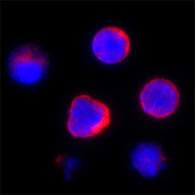
- Main image

- Experimental details
- Spi-B in Mouse Splenocytes. Spi-B was detected in immersion fixed mouse splenocytes using Sheep Anti-Mouse Spi-B Antigen Affinity-purified Polyclonal Antibody (Catalog # AF7204) at 15 µg/mL for 3 hours at room temperature. Cells were stained using the NorthernLights™ 557-conjugated Anti-Sheep IgG Secondary Antibody (red; Catalog # NL010) and counterstained with DAPI (blue). Specific staining was localized to plasma membranes and cytoplasm. View our protocol for Fluorescent ICC Staining of Non-adherent Cells.
Supportive validation
- Submitted by
- R&D Systems (provider)
- Main image
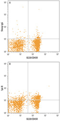
- Experimental details
- Detection of SPi-B in Mouse Splenocytes by Flow Cytometry. Mouse splenocytes were stained with Sheep Anti-Mouse Spi-B Antigen Affinity-purified Polyclonal Antibody (Catalog # AF7204) followed by Allophycocyanin-conjugated Anti-Sheep IgG Secondary Antibody (Catalog # F0127) and Rat Anti-Mouse B220/CD45R PE-conjugated Monoclonal Antibody (Catalog # FAB1217P). Quadrant markers were set based on control antibody staining (Catalog # 5-001-A). To facilitate intracellular staining, cells were fixed with paraformadehyde and permeabilized with saponin.
