 Explore
 Validate
 Learn
Learn Western blot
Antibody data
- Antibody Data
- Antigen structure
- References [0]
- Comments [0]
- Validations
- Western blot [2]
- Immunocytochemistry [1]
Submit
Validation data
Reference
Comment
Report error
- Product number
- OPA1-03170 - Provider product page

- Provider
- Invitrogen Antibodies
- Product name
- Anti-Phospho-EGFR (Tyr1068) Polyclonal Antibody
- Antibody type
- Polyclonal
- Antigen
- Synthetic peptide
- Description
- OPA1-03170 detects human phospho-EGFR (pTyr1068) from human samples. OPA1-03170 has been successfully used in Western blot procedures. By Western blot, this antibody detects an ~185 kDa protein representing phospho-EGFR (pTyr1068). OPA1-03170 immunizing phosphopeptide was derived from a region of human EGFR that contains tyrosine 1068.
- Reactivity
- Human
- Host
- Rabbit
- Isotype
- IgG
- Vial size
- 100 µL
- Storage
- -20° C, Avoid Freeze/Thaw Cycles
No comments: Submit comment
Supportive validation
- Submitted by
- Invitrogen Antibodies (provider)
- Main image
- Experimental details
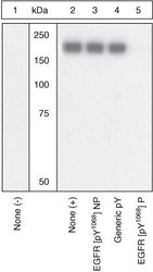
- Western blot analysis using a Phospho-EGFR pTyr1068 polyclonal antibody (Product # OPA1-03170).

- Submitted by
- Invitrogen Antibodies (provider)
- Main image

- Experimental details
- Western blot analysis was performed on whole cell extracts (30µg) of A-431 (Lane 1), A-431 treated with EGF (200 ng/mL for 10 minutes) (Lane2), A-431 treated with Afatinib followed by EGF (1uM of Afatinib for 6hrs, 200 ng/mL for 10 minutes) (Lane 3), A549 (Lane 4) and A549 treated with EGF (200 ng/mL for 10 minutes). The blots were probed with Phospho-EGFR (Tyr1068) Rabbit Polyclonal Antibody (Product# OPA1-03170, 1:500 dilution) and detected by chemiluminescence using Goat anti-Rabbit IgG (H+L) Superclonal™ Secondary Antibody, HRP conjugate (Product # A27036, 0.25µg/mL, 1:4000 dilution). A ~ 180 kDa band corresponding to Phospho-EGFR (Tyr1068) was increased upon EGF treatment across cell lines tested and pre-treatment with Afatinib (antagonist) resulted in inhibition of Phospho EGFR in A-431 cell line upon EGF treatment. Known quantity of protein samples were electrophoresed using Novex® NuPAGE® 4-12 % Bis-Tris gel (Product # NP0321BOX), XCell SureLock™ Electrophoresis System (Product # EI0002) and Novex® Sharp Pre-Stained Protein Standard (Product # LC5800). Resolved proteins were then transferred onto a nitrocellulose membrane by wet transfer method. The membrane was probed with the relevant primary and secondary Antibody following blocking with 5 % skimmed milk. Chemiluminescent detection was performed using Pierce™ ECL Western Blotting Substrate (Product # 32106).

Supportive validation
- Submitted by
- Invitrogen Antibodies (provider)
- Main image

- Experimental details
- Immunofluorescence analysis of Phospho-EGFR (Tyr1068) was performed using 70% confluent log phase A-431 cells treated with 200 ng/ml of EGF for 10 minutes. The cells were fixed with 4% paraformaldehyde for 10 minutes, permeabilized with 0.1% Triton™ X-100 for 10 minutes, and blocked with 1% BSA for 1 hour at room temperature. The cells were labeled with Phospho-EGFR (Tyr1068) Rabbit Polyclonal Antibody (Product# OPA1-03170) at 1:250 dilution in 0.1% BSA and incubated for 3 hours at room temperature and then labeled with Goat anti-Rabbit IgG (H+L) Superclonal™ Secondary Antibody, Alexa Fluor® 488 conjugate (A27034) at a dilution of 1:2000 for 45 minutes at room temperature (Panel a: green). Nuclei (Panel b: blue) were stained with SlowFade® Gold Antifade Mountant with DAPI (S36938). F-actin (Panel c: red) was stained with Rhodamine Phalloidin (Product # R415, 1:300). Panel d represents the merged image showing membrane localization. Panel e shows untreated cells with no signal. Panel f represents cells treated with antagonist, Afatinib (1µM for 6hrs) followed by EGF (200ng/ml for 10 minutes), showing no Phospho-EGFR staining. Panel g represents control cells with no primary antibody to assess background. The images were captured at 60X magnification.
