Explore
 Validate
 Learn
Learn Western blot
 Immunocytochemistry
Antibody data
- Antibody Data
- Antigen structure
- References [5]
- Comments [0]
- Validations
- Western blot [2]
- Immunohistochemistry [1]
- Flow cytometry [1]
Submit
Validation data
Reference
Comment
Report error
- Product number
- AF1095 - Provider product page

- Provider
- Novus Biologicals
- Product name
- Rabbit Polyclonal EGFR Antibody
- Antibody type
- Polyclonal
- Description
- Antigen Affinity-purified. Detects human EGFR when phosphorylated at Y1173 in Western blots.
- Reactivity
- Human
- Host
- Rabbit
- Conjugate
- Unconjugated
- Isotype
- IgG
- Vial size
- 50 ug
- Concentration
- LYOPH
- Storage
- Use a manual defrost freezer and avoid repeated freeze-thaw cycles. 12 months from date of receipt, -20 to -70 degreesC as supplied. 1 month, 2 to 8 degreesC under sterile conditions after reconstitution. 6 months, -20 to -70 degreesC under sterile conditions after reconstitution.
Submitted references ADAM17 substrate release in proximal tubule drives kidney fibrosis.
Phenotypic diversity of breast cancer-related mutations in metalloproteinase-disintegrin ADAM12.
Analysis of Epithelial Growth Factor-Receptor (EGFR) Phosphorylation in Uterine Smooth Muscle Tumors: Correlation to Mucin-1 and Galectin-3 Expression.
Protein C is an autocrine growth factor for human skin keratinocytes.
Repulsion of cerebellar granule neurons by chondroitin sulfate proteoglycans is mediated by MAPK pathway.
Kefaloyianni E, Muthu ML, Kaeppler J, Sun X, Sabbisetti V, Chalaris A, Rose-John S, Wong E, Sagi I, Waikar SS, Rennke H, Humphreys BD, Bonventre JV, Herrlich A
JCI insight 2016 Aug 18;1(13)
JCI insight 2016 Aug 18;1(13)
Phenotypic diversity of breast cancer-related mutations in metalloproteinase-disintegrin ADAM12.
Qi Y, Duhachek-Muggy S, Li H, Zolkiewska A
PloS one 2014;9(3):e92536
PloS one 2014;9(3):e92536
Analysis of Epithelial Growth Factor-Receptor (EGFR) Phosphorylation in Uterine Smooth Muscle Tumors: Correlation to Mucin-1 and Galectin-3 Expression.
Weissenbacher T, Vrekoussis T, Roeder D, Makrigiannakis A, Mayr D, Ditsch N, Friese K, Jeschke U, Dian D
International journal of molecular sciences 2013 Feb 28;14(3):4783-92
International journal of molecular sciences 2013 Feb 28;14(3):4783-92
Protein C is an autocrine growth factor for human skin keratinocytes.
Xue M, Campbell D, Jackson CJ
The Journal of biological chemistry 2007 May 4;282(18):13610-6
The Journal of biological chemistry 2007 May 4;282(18):13610-6
Repulsion of cerebellar granule neurons by chondroitin sulfate proteoglycans is mediated by MAPK pathway.
Kaneko M, Kubo T, Hata K, Yamaguchi A, Yamashita T
Neuroscience letters 2007 Aug 9;423(1):62-7
Neuroscience letters 2007 Aug 9;423(1):62-7
No comments: Submit comment
Supportive validation
- Submitted by
- Novus Biologicals (provider)
- Main image
- Experimental details
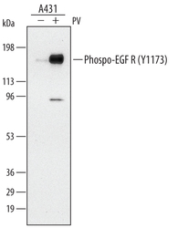
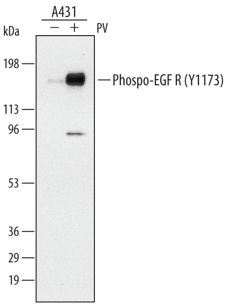
- Detection of Human Phospho-EGFR (Y1173) by Western Blot. Western blot shows lysates of A431 human epithelial carcinoma cell line untreated (-) or treated (+) with 100 μM pervanadate (PV) for 10 minutes. PVDF membrane was probed with 0.2 µg/mL of Rabbit Anti-Human Phospho-EGFR (Y1173) Antigen Affinity-purified Polyclonal Antibody, followed by HRP-conjugated Anti-Rabbit IgG Secondary Antibody (Catalog # HAF008). A specific band was detected for Phospho-EGFR (Y1173) at approximately 185 kDa (as indicated). This experiment was conducted under reducing conditions and using Immunoblot Buffer Group 1.
- Submitted by
- Novus Biologicals (provider)
- Main image
- Experimental details
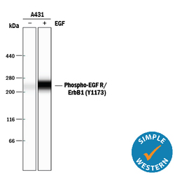
- Detection of Human Phospho-EGFR (Y1173) by Simple WesternTM. Simple Western lane view shows lysates of A431 human epithelial carcinoma cell line untreated (-) or treated (+) with 10 ng/mL Recombinant Human EGF (Catalog # 236-EG) for 5 minutes, loaded at 0.2 mg/mL. A specific band was detected for Phospho-EGFR (Y1173) at approximately 265 kDa (as indicated) using 2 µg/mL of Rabbit Anti-Human Phospho-EGFR (Y1173) Antigen Affinity-purified Polyclonal Antibody (Catalog # AF1095). This experiment was conducted under reducing conditions and using the 66-440 kDa separation system.

Supportive validation
- Submitted by
- Novus Biologicals (provider)
- Main image

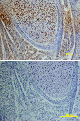
- Experimental details
- Phospho-EGFR (Y1173) in Mouse Embryo. EGFR phosphorylated at Y1173 was detected in immersion fixed frozen sections of mouse embryo using Rabbit Anti-Human Phospho-EGFR (Y1173) Antigen Affinity-purified Polyclonal Antibody (Catalog # AF1095) at 15 µg/mL overnight at 4 °C. Tissue was stained using the Anti-Rabbit HRP-DAB Cell & Tissue Staining Kit (brown; Catalog # CTS005) and counterstained with hematoxylin (blue). Lower panel shows a lack of labeling if primary antibodies are omitted and tissue is stained only with secondary antibody followed by incubation with detection reagents. View our protocol for Chromogenic IHC Staining of Frozen Tissue Sections.
Supportive validation
- Submitted by
- Novus Biologicals (provider)
- Main image

- Experimental details
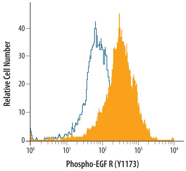
- Detection of Phospho-EGFR (Y1173) in A431 Human Cell Line by Flow Cytometry. A431 human epithelial carcinoma cells were untreated (open histogram), or treated for 5 minutes with 100 ng/mL Recombinant Human EGF (Catalog # 236-EG; filled histogram) then stained with Rabbit Anti-Human Phospho-EGFR (Y1173) Antigen Affinity-purified Polyclonal Antibody (Catalog # AF1095), followed by Phycoerythrin-conjugated Anti-Rabbit IgG Secondary Antibody (Catalog # F0110). To facilitate intracellular staining, cells were fixed with para-formaldehyde and permeabilized with saponin.