Explore
 Validate
 Learn
Learn Western blot
 Immunocytochemistry
Antibody data
- Antibody Data
- Antigen structure
- References [1]
- Comments [0]
- Validations
- Immunocytochemistry [1]
- Flow cytometry [1]
Submit
Validation data
Reference
Comment
Report error
- Product number
- 701053 - Provider product page

- Provider
- Invitrogen Antibodies
- Product name
- Anti-Phospho-EGFR (Tyr1068) Antibody (2H9L3), ABfinity™ Rabbit Monoclonal
- Antibody type
- Monoclonal
- Antigen
- Synthetic peptide
- Description
- This antibody is predicted to react with human based on sequence homology. ABfinity recombinant antibodies are rabbit monoclonal antibodies, unmatched for producing superior results. ABfinity antibodies are developed by immunizing animals, screening for functionality, cloning the immunogen-specific antibody genes into high-level mammalian expression vectors, produced on a large scale, and purified with Protein A. ABfinity monoclonal antibodies resemble rabbit monoclonals isolated from serum or produced by hybridomas, but demonstrate greater specificity and sensitivity. Because ABfinity recombinant antibodies are derived from cloned DNA sequences of the heavy and light antibody chains, they are not susceptible to cell-line drift or lot-to-lot variation, thus allowing for peak specificity and performance. Intact IgG appears on a non-reducing gel as ~150 kDa band and upon reduction generating a ~25 kDa light chain band and a ~50 kDa heavy chain.
- Reactivity
- Human
- Host
- Rabbit
- Isotype
- IgG
- Antibody clone number
- 2H9L3
- Vial size
- 100 µg
- Concentration
- 0.5 mg/ml
- Storage
- Maintain refrigerated at 2-8°C for up to 1 month. For long term storage store at -20°C
Submitted references Evaluation of dimerization-inhibitory activities of cyclic peptides containing a β-hairpin loop sequence of the EGF receptor.
Mizuguchi T, Ohara N, Iida M, Ninomiya R, Wada S, Kiso Y, Saito K, Akaji K
Bioorganic & medicinal chemistry 2012 Oct 1;20(19):5730-7
Bioorganic & medicinal chemistry 2012 Oct 1;20(19):5730-7
No comments: Submit comment
Supportive validation
- Submitted by
- Invitrogen Antibodies (provider)
- Main image

- Experimental details
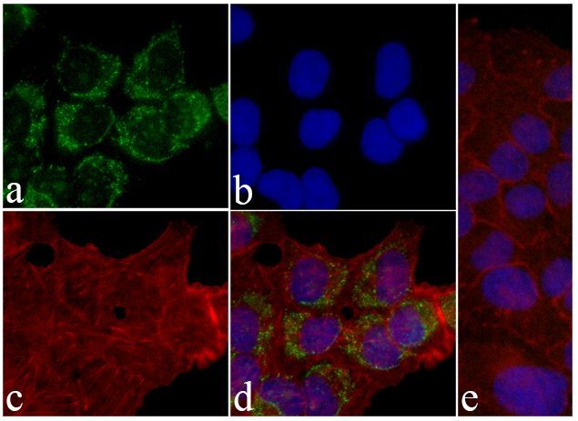
- Immunofluorescent analysis of EGFR (pY1068) was performed on 70% confluent log phase A549 cells. The cells were fixed with 4% paraformaldehyde for 15 minutes, permeabilized with 0. 25% Triton X-100 for 10 minutes, and blocked with 5% BSA for 1 hour at room temperature. The cells were labeled with ABfinity EGFR (pY1068) recombinant rabbit monoclonal antibody (Product # 701053) at a dilution of 1:500 in 1% BSA and incubated for 3 hours at room temperature and then labeled with Alexa Fluor® 488 goat anti-rabbit IgG secondary antibody (Product # A-11008) at a dilution of 1:400 for 30 minutes at room temperature (Panel a: green). Nuclei (Panel b: blue) were stained with SlowFade® Gold Antifade Mountant DAPI (Product # S36938). F-actin (Panel c: red) was stained with Alexa Fluor® 594 phalloidin (Product # A12381). Panel d is a merged image showing cytoplasmic localization of phosphorylated EGFR (pY1068). Panel e shows competition with the phospho-AKT (pT308) peptide. The images were captured using a Nikon microscope at 20X magnification.
Supportive validation
- Submitted by
- Invitrogen Antibodies (provider)
- Main image

- Experimental details
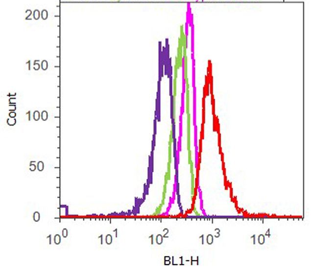
- Flow cytometry analysis of EGFR (pY1068) was performed on A549 cells treated with EGF (100 ng/ml, 5 minutes). Cells were fixed with 70% ethanol for 10 minutes, permeabilized with 0. 25% Tritonª X-100 for 20 minutes, and blocked with 5% BSA for 1 hour at room temperature. Cells were labeled with ABfinityª EGFR (pY1068) recombinant rabbit monoclonal antibody (Product # 701053, red histogram) or with rabbit isotype control (pink histogram) at a dilution of 1:250 in 2.5% BSA. After incubation at room temperature for 3 hours, the cells were labeled with Alexa Fluor¨ 488 goat anti-Rabbit Secondary antibody (Product # A11008) at a dilution of 1:400 for 30 minutes at room temperature. The representative 10,000 cells were acquired and analyzed for each sample using an Attune¨ Acoustic Focusing Cytometer. The purple histogram represents unstained control cells and the green histogram represents no-primary-antibody control.