Explore
 Validate
 Learn
Learn Western blot
Antibody data
- Antibody Data
- Antigen structure
- References [4]
- Comments [0]
- Validations
- Western blot [2]
- Immunocytochemistry [1]
- Flow cytometry [1]
- Other assay [1]
Submit
Validation data
Reference
Comment
Report error
- Product number
- 44-792G - Provider product page

- Provider
- Invitrogen Antibodies
- Product name
- Phospho-EGFR (Tyr1148) Polyclonal Antibody
- Antibody type
- Polyclonal
- Antigen
- Synthetic peptide
- Reactivity
- Human
- Host
- Rabbit
- Isotype
- IgG
- Vial size
- 100 µL
- Storage
- -20°C
Submitted references Time-resolved multimodal analysis of Src Homology 2 (SH2) domain binding in signaling by receptor tyrosine kinases.
A portrait of tissue phosphoprotein stability in the clinical tissue procurement process.
Enhanced activation of epidermal growth factor receptor caused by tumor-derived E-cadherin mutations.
Aplidin induces apoptosis in human cancer cells via glutathione depletion and sustained activation of the epidermal growth factor receptor, Src, JNK, and p38 MAPK.
Jadwin JA, Oh D, Curran TG, Ogiue-Ikeda M, Jia L, White FM, Machida K, Yu J, Mayer BJ
eLife 2016 Apr 12;5:e11835
eLife 2016 Apr 12;5:e11835
A portrait of tissue phosphoprotein stability in the clinical tissue procurement process.
Espina V, Edmiston KH, Heiby M, Pierobon M, Sciro M, Merritt B, Banks S, Deng J, VanMeter AJ, Geho DH, Pastore L, Sennesh J, Petricoin EF 3rd, Liotta LA
Molecular & cellular proteomics : MCP 2008 Oct;7(10):1998-2018
Molecular & cellular proteomics : MCP 2008 Oct;7(10):1998-2018
Enhanced activation of epidermal growth factor receptor caused by tumor-derived E-cadherin mutations.
Bremm A, Walch A, Fuchs M, Mages J, Duyster J, Keller G, Hermannstädter C, Becker KF, Rauser S, Langer R, von Weyhern CH, Höfler H, Luber B
Cancer research 2008 Feb 1;68(3):707-14
Cancer research 2008 Feb 1;68(3):707-14
Aplidin induces apoptosis in human cancer cells via glutathione depletion and sustained activation of the epidermal growth factor receptor, Src, JNK, and p38 MAPK.
Cuadrado A, Garcia-Fernandez LF, Gonzalez L, Suarez Y, Losada A, Alcaide V, Martinez T, Fernandez-Sousa JM, Sanchez-Puelles JM, Munoz A
The Journal of biological chemistry 2003 Jan 3;278(1):241-50
The Journal of biological chemistry 2003 Jan 3;278(1):241-50
No comments: Submit comment
Supportive validation
- Submitted by
- Invitrogen Antibodies (provider)
- Main image
- Experimental details
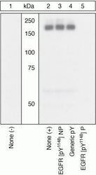
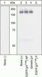
- Peptide Competition. Extracts of A431 cells unstimulated (1) or stimulated with 200 ng/mL EGF (2-5) for 15 minutes were resolved by SDS-PAGE on a 10% Tris-glycine gel and transferred to PVDF. The membrane was blocked with a 5% BSA-TBST buffer for one hour at room temperature, then incubated with the EGFR [pY1148] antibody for two hours at room temperature in a 1% BSA-TBST buffer, following prior incubation with: no peptide (1, 2), non-phosphopeptide corresponding to the phosphopeptide immunogen (3), a generic phosphotyrosine-containing peptide (4), or the phosphopeptide immunogen (5). After washing, the membrane was incubated with goat F(ab’)2 anti-rabbit IgG HRP conjugate (Product # ALI4404) and signals were detected using the Pierce SuperSignal™ method. The data show that only the phosphopeptide corresponding to EGFR [pY1148] blocks the signal, demonstrating the specificity of the antibody. The data also show the induction of EGFR [pY1148] phosphorylation by the addition of EGF to this cell system.
- Submitted by
- Invitrogen Antibodies (provider)
- Main image
- Experimental details
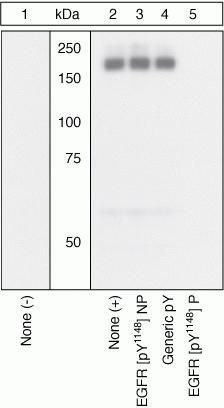
- Peptide Competition. Extracts of A431 cells unstimulated (1) or stimulated with 200 ng/mL EGF (2-5) for 15 minutes were resolved by SDS-PAGE on a 10% Tris-glycine gel and transferred to PVDF. The membrane was blocked with a 5% BSA-TBST buffer for one hour at room temperature, then incubated with the EGFR [pY1148] antibody for two hours at room temperature in a 1% BSA-TBST buffer, following prior incubation with: no peptide (1, 2), non-phosphopeptide corresponding to the phosphopeptide immunogen (3), a generic phosphotyrosine-containing peptide (4), or the phosphopeptide immunogen (5). After washing, the membrane was incubated with goat F(ab’)2 anti-rabbit IgG HRP conjugate (Product # ALI4404) and signals were detected using the Pierce SuperSignal™ method. The data show that only the phosphopeptide corresponding to EGFR [pY1148] blocks the signal, demonstrating the specificity of the antibody. The data also show the induction of EGFR [pY1148] phosphorylation by the addition of EGF to this cell system.

Supportive validation
- Submitted by
- Invitrogen Antibodies (provider)
- Main image
- Experimental details
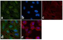
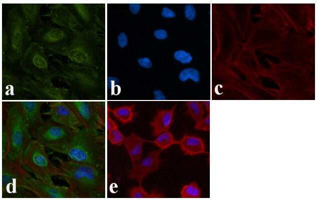
- Immunofluorescence analysis of EGFR (pY1148) was done on 70% confluent log phase A549. The cells were fixed with 4% paraformaldehyde for 15 minutes, permeabilized with 0.25% Triton™ X-100 for 10 minutes, and blocked with 5% BSA for 1 hour at room temperature. The cells were labeled with EGFR (pY1148) Rabbit polyclonal Antibody (Product # 44-792G) at 1:250 dilution in 1% BSA and incubated for 3 hours at room temperature and then labeled with Alexa Fluor 488 Goat Anti-Rabbit IgG Secondary Antibody (Product # A-11008) at a dilution of 1:400 for 30 minutes at room temperature (Panel a: green). Nuclei (Panel b: blue) were stained with SlowFade® Gold Antifade Mountant DAPI (Product # S36938). F-actin (Panel c: red) was stained with Alexa Fluor 594 Phalloidin (Product # A12381). Panel d is a merged image showing cytoplasmic localization. Panel e shows no primary antibody control. The images were captured at 20X magnification.
Supportive validation
- Submitted by
- Invitrogen Antibodies (provider)
- Main image
- Experimental details
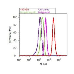
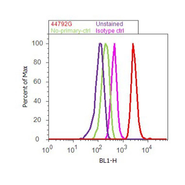
- Flow cytometry analysis of EGFR [pY1148] was done on A549 cells treated with EGF (200ng/ml, 10 minutes). Cells were fixed with 70% ethanol for 10 minutes, permeabilized with 0.25% Tritonª X-100 for 20 minutes, and blocked with 5% BSA for 30 minutes at room temperature. Cells were labeled with EGFR [pY1148] Rabbit Polyclonal Antibody (44792G, red histogram) or with rabbit isotype control (pink histogram) at 3-5 µg/million cells in 2.5% BSA. After incubation at room temperature for 2 hours, the cells were labeled with Alexa Fluor¨ 488 Goat Anti-Rabbit Secondary Antibody (A11008) at a dilution of 1:400 for 30 minutes at room temperature. The representative 10,000 cells were acquired and analyzed for each sample using an Attune¨ Acoustic Focusing Cytometer. The purple histogram represents unstained control cells and the green histogram represents no-primary-antibody control.
Supportive validation
- Submitted by
- Invitrogen Antibodies (provider)
- Main image
- Experimental details
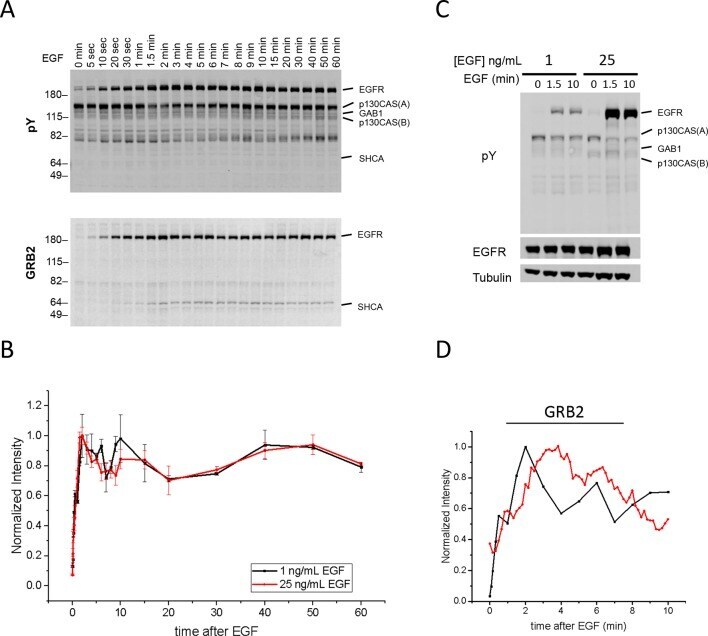
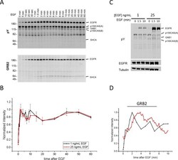
- Figure 4--figure supplement 6. Quantification of Grb2 binding sites and in vivo Grb2 SH2 recruitment in A431 cells stimulated with 1 ng/ml EGF. ( A ) Representative anti-pY immunoblot (upper panel) and Grb2 SH2 far-western blot (lower panel) of A431 cells stimulated with 1 ng/mL EGF and flash frozen at 22 discrete time points. ( B ) Quantification of EGFR tyrosine phosphorylation kinetics (from anti-pY immunoblot) in A431 cells treated with 1 ng/mL EGF (n=2 biological replicates) and 25 ng/mL EGF (n=3 biological replicates). ( C ) Anti-pY immunoblot of A431 cells stimulated with 1 ng/mL and 25 ng/mL EGF at 0, 1.5 and 10 min. EGFR phosphorylation was 5.4 +/- 0.4 fold greater in cells stimulated with 25 ng/mL (normalized for EGFR expression, error = SEM). No difference was observed in prestimulation EGFR phosphorylation after normalization. ( D ) Comparison of Grb2 SH2 binding site phosphorylation kinetics (GST-Grb2 SH2 FW, black; time constant tau = 54.6 +/- 1.4 s, n=2 biological replicates) and Grb2 SH2 in vivo membrane recruitment kinetics (tdEOS-GRB2 SH2 TIRF, red; time constant tau = 116.7 +/- 2.3 s, n=2 biological replicates). DOI: http://dx.doi.org/