Explore
 Validate
 Learn
Learn Western blot
 Flow cytometry
Antibody data
- Antibody Data
- Antigen structure
- References [4]
- Comments [0]
- Validations
- Flow cytometry [2]
Submit
Validation data
Reference
Comment
Report error
- Product number
- NBP2-50599 - Provider product page

- Provider
- Novus Biologicals
- Product name
- Mouse Monoclonal EGFR Antibody
- Antibody type
- Monoclonal
- Description
- Protein G purified. Clone DH8.3 of EGF R/ErbB1 antibody is specific for Truncated EGFR which is also called EGFR type III, EGFRvIII or delta-EGFR (characterised by a deletion of 267 amino acids in the extracellular domain, leading to a receptor which is unable to bind ligand yet is constitutively active). Because of its impaired internalisation and degradation, EGFRvIII is known to enhance tumourigenesis. NOTE: Clone DH8.3 DOES NOT cross-react with full length or wildtype EGFR (wtEGFR).
- Reactivity
- Human
- Host
- Mouse
- Isotype
- IgG
- Vial size
- 0.1 ml
- Concentration
- 1 mg/ml
- Storage
- Store at 4C short term. Aliquot and store at -20C long term. Avoid freeze-thaw cycles.
Submitted references Poly[N-(2-hydroxypropyl)methacrylamide]-Modified Magnetic γ-F2 O3 Nanoparticles Conjugated with Doxorubicin for Glioblastoma Treatment.
Immunohistochemical analysis of the mutant epidermal growth factor, deltaEGFR, in glioblastoma.
A monoclonal antibody recognizing human cancers with amplification/overexpression of the human epidermal growth factor receptor.
Novel monoclonal antibody specific for the de2-7 epidermal growth factor receptor (EGFR) that also recognizes the EGFR expressed in cells containing amplification of the EGFR gene.
Plichta Z, Horák D, Mareková D, Turnovcová K, Kaiser R, Jendelová P
ChemMedChem 2020 Jan 7;15(1):96-104
ChemMedChem 2020 Jan 7;15(1):96-104
Immunohistochemical analysis of the mutant epidermal growth factor, deltaEGFR, in glioblastoma.
Nishikawa R, Sugiyama T, Narita Y, Furnari F, Cavenee WK, Matsutani M
Brain tumor pathology 2004;21(2):53-6
Brain tumor pathology 2004;21(2):53-6
A monoclonal antibody recognizing human cancers with amplification/overexpression of the human epidermal growth factor receptor.
Jungbluth AA, Stockert E, Huang HJ, Collins VP, Coplan K, Iversen K, Kolb D, Johns TJ, Scott AM, Gullick WJ, Ritter G, Cohen L, Scanlan MJ, Cavenee WK, Old LJ
Proceedings of the National Academy of Sciences of the United States of America 2003 Jan 21;100(2):639-44
Proceedings of the National Academy of Sciences of the United States of America 2003 Jan 21;100(2):639-44
Novel monoclonal antibody specific for the de2-7 epidermal growth factor receptor (EGFR) that also recognizes the EGFR expressed in cells containing amplification of the EGFR gene.
Johns TG, Stockert E, Ritter G, Jungbluth AA, Huang HJ, Cavenee WK, Smyth FE, Hall CM, Watson N, Nice EC, Gullick WJ, Old LJ, Burgess AW, Scott AM
International journal of cancer 2002 Mar 20;98(3):398-408
International journal of cancer 2002 Mar 20;98(3):398-408
No comments: Submit comment
Supportive validation
- Submitted by
- Novus Biologicals (provider)
- Main image
- Experimental details
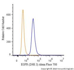
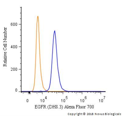
- Flow Cytometry: EGFR Antibody (DH8.3) - EGFRvIII [NBP2-50599] - An intracellular stain was performed on A431 cells with EGFR [DH8.3] Antibody NBP2-50599AF700 (blue) and a matched isotype control (orange). Cells were fixed with 4% PFA and then permeabilized with 0.1% saponin. Cells were incubated in an antibody dilution of 5 ug/mL for 30 minutes at room temperature. Both antibodies were conjugated to Alexa Fluor 700.
- Submitted by
- Novus Biologicals (provider)
- Main image
- Experimental details
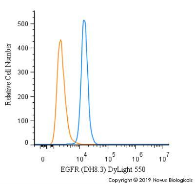
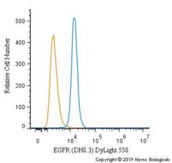
- Flow Cytometry: EGFR Antibody (DH8.3) - EGFRvIII [NBP2-50599] - An intracellular stain was performed on A431 cells with EGFR [DH8.3] Antibody NBP2-50599R (blue) and a matched isotype control (orange). Cells were fixed with 4% PFA and then permeabilized with 0.1% saponin. Cells were incubated in an antibody dilution of 5 ug/mL for 30 minutes at room temperature. Both antibodies were conjugated to DyLight 550.