Explore
 Validate
 Learn
Learn Immunocytochemistry
Antibody data
- Antibody Data
- Antigen structure
- References [5]
- Comments [0]
- Validations
- Immunocytochemistry [1]
- Flow cytometry [1]
Submit
Validation data
Reference
Comment
Report error
- Product number
- 50-9509-41 - Provider product page

- Provider
- Invitrogen Antibodies
- Product name
- EGFR Monoclonal Antibody (me1B3), eFluor™ 660, eBioscience™
- Antibody type
- Monoclonal
- Antigen
- Other
- Description
- Description: This me1B3 monoclonal antibody reacts with human epidermal growth factor receptor (EGFR), also known as ErbB1 and Her1. EGFR is a receptor tyrosine kinase belonging to the EGFR/ErbB receptor subfamily, which also includes ErbB2/Her2/neu. EGFR contains an extracellular ligand-binding domain, a single transmembrane-spanning region, and a cytoplasmic tail containing a tyrosine kinase domain. Specific ligands include EGF and TNF alpha, as well as other members of the EGF protein family. Upon ligand binding, EGFR undergoes receptor-mediated dimerization with itself or ErbB2/Her2/neu. Dimerization induces auto-phosphorylation, resulting in the activation of PLC gamma, JAK/STAT, MAPK/ERK, and PI3K/AKT signaling pathways. EGFR has a role in cell proliferation, migration and differentiation, and is evolutionarily highly conserved in structure and function. EGFR is broadly expressed and is overexpressed or mutated in glioblastomas and a number of different epithelial cancers, including non-small cell lung cancer and colorectal cancer. Applications Reported: This me1B3 antibody has been reported for use in flow cytometric analysis. Applications Tested: This me1B3 antibody has been pre-titrated and tested by flow cytometric analysis of HeLa cells. This can be used at 5 µL (0.125 µg) per test. A test is defined as the amount (µg) of antibody that will stain a cell sample in a final volume of 100 µL. Cell number should be determined empirically but can range from 10^5 to 10^8 cells/test. eFluor™ 660 is a replacement for Alexa Fluor™ 647. eFluor™ 660 emits at 659 nm and is excited with the red laser (633 nm). Please make sure that your instrument is capable of detecting this fluorochome. Excitation: 633-647 nm; Emission: 668 nm; Laser: Red Laser. Filtration: 0.2 µm post-manufacturing filtered.
- Reactivity
- Human
- Host
- Mouse
- Isotype
- IgG
- Antibody clone number
- me1B3
- Vial size
- 25 Tests
- Concentration
- 5 μL/Test
- Storage
- 4°C, store in dark, DO NOT FREEZE!
Submitted references Oncogenic mutations counteract intrinsic disorder in the EGFR kinase and promote receptor dimerization.
Cell signaling by receptor tyrosine kinases.
Crystal structure of human epidermal growth factor and its dimerization.
Amplification, enhanced expression and possible rearrangement of EGF receptor gene in primary human brain tumours of glial origin.
Amplification and enhanced expression of the epidermal growth factor receptor gene in A431 human carcinoma cells.
Shan Y, Eastwood MP, Zhang X, Kim ET, Arkhipov A, Dror RO, Jumper J, Kuriyan J, Shaw DE
Cell 2012 May 11;149(4):860-70
Cell 2012 May 11;149(4):860-70
Cell signaling by receptor tyrosine kinases.
Lemmon MA, Schlessinger J
Cell 2010 Jun 25;141(7):1117-34
Cell 2010 Jun 25;141(7):1117-34
Crystal structure of human epidermal growth factor and its dimerization.
Lu HS, Chai JJ, Li M, Huang BR, He CH, Bi RC
The Journal of biological chemistry 2001 Sep 14;276(37):34913-7
The Journal of biological chemistry 2001 Sep 14;276(37):34913-7
Amplification, enhanced expression and possible rearrangement of EGF receptor gene in primary human brain tumours of glial origin.
Libermann TA, Nusbaum HR, Razon N, Kris R, Lax I, Soreq H, Whittle N, Waterfield MD, Ullrich A, Schlessinger J
Nature 1985 Jan 10-18;313(5998):144-7
Nature 1985 Jan 10-18;313(5998):144-7
Amplification and enhanced expression of the epidermal growth factor receptor gene in A431 human carcinoma cells.
Merlino GT, Xu YH, Ishii S, Clark AJ, Semba K, Toyoshima K, Yamamoto T, Pastan I
Science (New York, N.Y.) 1984 Apr 27;224(4647):417-9
Science (New York, N.Y.) 1984 Apr 27;224(4647):417-9
No comments: Submit comment
Supportive validation
- Submitted by
- Invitrogen Antibodies (provider)
- Main image
- Experimental details
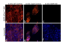
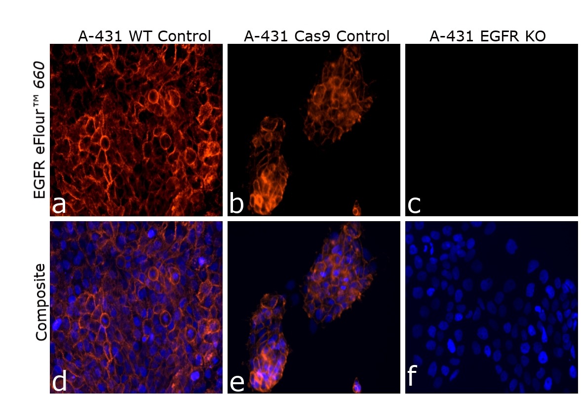
- Knockout of EGFR was achieved by CRISPR-Cas9 genome editing using LentiArray™ Lentiviral sgRNA (Product # A32042) and LentiArray Cas9 Lentivirus (Product # A32064). Immunofluorescence analysis was performed on wild type A-431 cells (panel a,d), A-431 Cas9 control cells (panel b,e) and A-431 EGFRKO cells (panel c, f). Cells were fixed, permeabilized, and labeled with EGFR Monoclonal Antibody (me1B3), eFluor™ 660, eBioscience™(Product # 50-9509-42, 1:50). Nuclei (blue) were stained using ProLong™ Diamond Antifade Mountant with DAPI (Product # P36962). Loss of signal (panel c,f) upon CRISPR mediated knockout (KO) confirms that antibody is specific to NF-H (green). The images were captured at 20X magnification.
Supportive validation
- Submitted by
- Invitrogen Antibodies (provider)
- Main image
- Experimental details
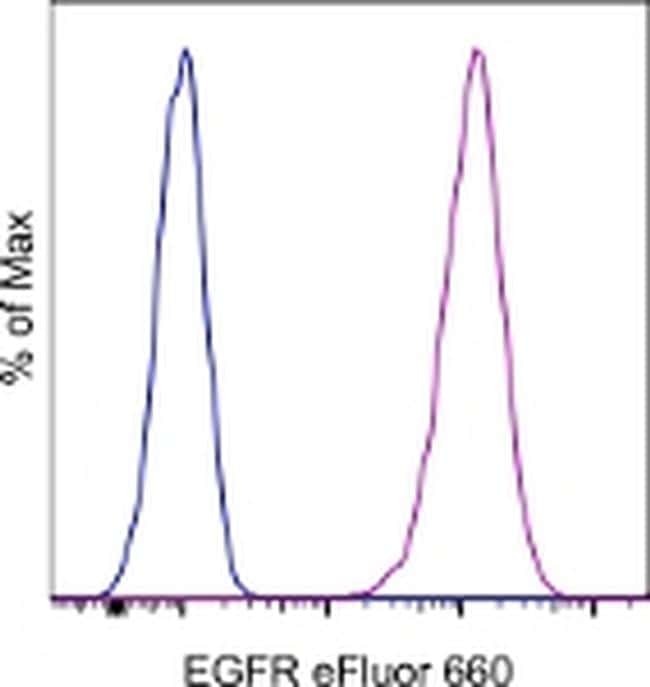
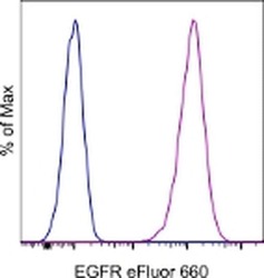
- Staining of HeLa cells with Mouse IgG1 K Isotype Control eFluor® 660 (Product # 50-4714-82) (blue histogram) or Anti-Human Epidermal Growth Factor Receptor (EGFR) eFluor® 660 (purple histogram). Total viable cells, as determined by Fixable Viability Dye eFluor® 506 (Product # 65-0866-14), were used for analysis.