Explore
 Validate
 Learn
Learn Western blot
 ELISA
ELISAAntibody data
- Antibody Data
- Antigen structure
- References [6]
- Comments [0]
- Validations
- Western blot [1]
Submit
Validation data
Reference
Comment
Report error
- Product number
- A00023-4 - Provider product page

- Provider
- Boster Biological Technology
- Product name
- Anti-EGFR Antibody Picoband™
- Antibody type
- Polyclonal
- Description
- Rabbit IgG polyclonal antibody for EGFR detection. Tested with WB, IHC-P, ICC/IF, IF, FCM, Direct ELISA in Human;Mouse;Rat.
- Reactivity
- Human, Mouse, Rat
- Host
- Rabbit
- Vial size
- 100μg/vial
- Concentration
- Add 0.2ml of distilled water will yield a concentration of 500ug/ml.
- Storage
- At -20°C for one year. After reconstitution, at 4°C for one month. It can also be aliquoted and stored frozen at -20°C for a longer time. Avoid repeated freezing and thawing.
- Handling
- Add 0.2ml of distilled water will yield a concentration of 500ug/ml.
Submitted references A microfluidic lung-on-a-chip based on biomimetic hydrogel membrane.
Circular RNA encoded MET variant promotes glioblastoma tumorigenesis.
Curcumin derivative L6H4 inhibits proliferation and invasion of gastric cancer cell line BGC-823.
Localization of epidermal growth factor (EGF) and its receptor (EGFR) during postnatal testis development in the alpaca (Lama pacos).
Epidermal growth factor-induced proliferation of chicken primordial germ cells: involvement of calcium/protein kinase C and NFKB1.
Interactive actions of prostaglandin and epidermal growth factor to enhance proliferation of granulosa cells from chicken prehierarchical follicles.
Shen C, Yang H, She W, Meng Q
Biotechnology and bioengineering 2023 Jul;120(7):2027-2038
Biotechnology and bioengineering 2023 Jul;120(7):2027-2038
Circular RNA encoded MET variant promotes glioblastoma tumorigenesis.
Zhong J, Wu X, Gao Y, Chen J, Zhang M, Zhou H, Yang J, Xiao F, Yang X, Huang N, Qi H, Wang X, Bai F, Shi Y, Zhang N
Nature communications 2023 Jul 25;14(1):4467
Nature communications 2023 Jul 25;14(1):4467
Curcumin derivative L6H4 inhibits proliferation and invasion of gastric cancer cell line BGC-823.
Mu J, Wang X, Dong L, Sun P
Journal of cellular biochemistry 2019 Jan;120(1):1011-1017
Journal of cellular biochemistry 2019 Jan;120(1):1011-1017
Localization of epidermal growth factor (EGF) and its receptor (EGFR) during postnatal testis development in the alpaca (Lama pacos).
He J, Dong C, You R, Zhu Z, Lv L, Smith GW
Animal reproduction science 2009 Nov;116(1-2):155-61
Animal reproduction science 2009 Nov;116(1-2):155-61
Epidermal growth factor-induced proliferation of chicken primordial germ cells: involvement of calcium/protein kinase C and NFKB1.
Ge C, Yu M, Petitte JN, Zhang C
Biology of reproduction 2009 Mar;80(3):528-36
Biology of reproduction 2009 Mar;80(3):528-36
Interactive actions of prostaglandin and epidermal growth factor to enhance proliferation of granulosa cells from chicken prehierarchical follicles.
Jin Y, Zhang C, Zeng W, Taya K, Tan TQ
Prostaglandins & other lipid mediators 2007 Jun;83(4):285-94
Prostaglandins & other lipid mediators 2007 Jun;83(4):285-94
No comments: Submit comment
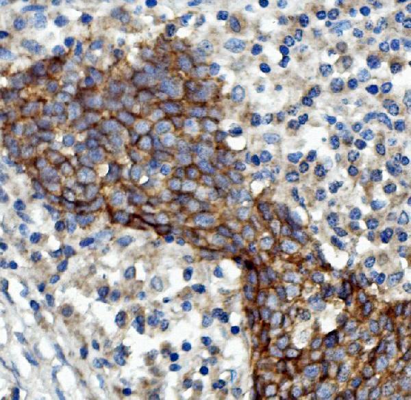
Supportive validation
- Submitted by
- Boster Biological Technology (provider)
- Main image
- Experimental details
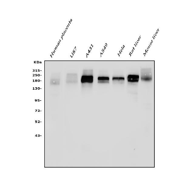
- Western blot analysis of EGFR using anti-EGFR antibody (A00023-4). Electrophoresis was performed on a 5-20% SDS-PAGE gel at 70V (Stacking gel) / 90V (Resolving gel) for 2-3 hours. The sample well of each lane was loaded with 50ug of sample under reducing conditions. Lane 1: human placenta tissue lysates, Lane 2: human U87 whole cell lysates, Lane 3: human A431 whole cell lysates, Lane 4: human A549 whole cell lysates, Lane 5: human Hela whole cell lysates, Lane 6: rat liver tissue lysates, Lane 7: mouse liver tissue lysates. After Electrophoresis, proteins were transferred to a Nitrocellulose membrane at 150mA for 50-90 minutes. Blocked the membrane with 5% Non-fat Milk/ TBS for 1.5 hour at RT. The membrane was incubated with rabbit anti-EGFR antigen affinity purified polyclonal antibody (Catalog # A00023-4) at 0.25 μg/mL overnight at 4°C, then washed with TBS-0.1%Tween 3 times with 5 minutes each and probed with a goat anti-rabbit IgG-HRP secondary antibody at a dilution of 1:10000 for 1.5 hour at RT. The signal is developed using an Enhanced Chemiluminescent detection (ECL) kit (Catalog # EK1002) with Tanon 5200 system. A specific band was detected for EGFR at approximately 180KD. The expected band size for EGFR is at 180KD.
- Additional image