Explore
 Validate
 Learn
Learn Western blot
 ELISA
ELISAAntibody data
- Antibody Data
- Antigen structure
- References [4]
- Comments [0]
- Validations
- Western blot [1]
Submit
Validation data
Reference
Comment
Report error
- Product number
- A00023-5 - Provider product page

- Provider
- Boster Biological Technology
- Product name
- Anti-EGFR Antibody Picoband™
- Antibody type
- Polyclonal
- Description
- Rabbit IgG polyclonal antibody for EGFR detection. Tested with WB, IHC-P, ICC/IF, FCM, Direct ELISA in Human.
- Reactivity
- Human
- Host
- Rabbit
- Vial size
- 100μg/vial
- Concentration
- Add 0.2ml of distilled water will yield a concentration of 500ug/ml.
- Storage
- At -20°C for one year. After reconstitution, at 4°C for one month. It can also be aliquoted and stored frozen at -20°C for a longer time. Avoid repeated freezing and thawing.
- Handling
- Add 0.2ml of distilled water will yield a concentration of 500ug/ml.
Submitted references A microfluidic lung-on-a-chip based on biomimetic hydrogel membrane.
Microbiota, co-metabolites, and network pharmacology reveal the alteration of the ginsenoside fraction on inflammatory bowel disease.
Inhibition of angiogenesis involves in anticancer activity of riccardin D, a macrocyclic bisbibenzyl, in human lung carcinoma.
Epidermal growth factor receptor in cultured human retinal pigment epithelial cells.
Shen C, Yang H, She W, Meng Q
Biotechnology and bioengineering 2023 Jul;120(7):2027-2038
Biotechnology and bioengineering 2023 Jul;120(7):2027-2038
Microbiota, co-metabolites, and network pharmacology reveal the alteration of the ginsenoside fraction on inflammatory bowel disease.
Wang D, Guo M, Li X, Zhao D, Wang M
Journal of ginseng research 2023 Jan;47(1):54-64
Journal of ginseng research 2023 Jan;47(1):54-64
Inhibition of angiogenesis involves in anticancer activity of riccardin D, a macrocyclic bisbibenzyl, in human lung carcinoma.
Sun CC, Zhang YS, Xue X, Cheng YN, Liu HP, Zhao CR, Lou HX, Qu XJ
European journal of pharmacology 2011 Sep 30;667(1-3):136-43
European journal of pharmacology 2011 Sep 30;667(1-3):136-43
Epidermal growth factor receptor in cultured human retinal pigment epithelial cells.
Yan F, Hui YN, Li YJ, Guo CM, Meng H
Ophthalmologica. Journal international d'ophtalmologie. International journal of ophthalmology. Zeitschrift fur Augenheilkunde 2007;221(4):244-50
Ophthalmologica. Journal international d'ophtalmologie. International journal of ophthalmology. Zeitschrift fur Augenheilkunde 2007;221(4):244-50
No comments: Submit comment
Supportive validation
- Submitted by
- Boster Biological Technology (provider)
- Main image
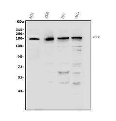
- Experimental details
- Western blot analysis of EGFR using anti-EGFR antibody (A00023-5). Electrophoresis was performed on a 5-20% SDS-PAGE gel at 70V (Stacking gel) / 90V (Resolving gel) for 2-3 hours. The sample well of each lane was loaded with 50ug of sample under reducing conditions. Lane 1: human A431 whole cell lysates, Lane 2: human A549 whole cell lysates, Lane 3: human U87 whole cell lysates, Lane 4: human HELA whole cell lysates. After Electrophoresis, proteins were transferred to a Nitrocellulose membrane at 150mA for 50-90 minutes. Blocked the membrane with 5% Non-fat Milk/ TBS for 1.5 hour at RT. The membrane was incubated with rabbit anti-EGFR antigen affinity purified polyclonal antibody (Catalog # A00023-5) at 0.5 μg/mL overnight at 4°C, then washed with TBS-0.1%Tween 3 times with 5 minutes each and probed with a goat anti-rabbit IgG-HRP secondary antibody at a dilution of 1:5000 for 1.5 hour at RT. The signal is developed using an Enhanced Chemiluminescent detection (ECL) kit (Catalog # EK1002) with Tanon 5200 system. A specific band was detected for EGFR at approximately 180KD. The expected band size for EGFR is at 134KD.
- Additional image