Explore
 Validate
 Learn
Learn Western blot
Antibody data
- Antibody Data
- Antigen structure
- References [3]
- Comments [0]
- Validations
- Western blot [1]
- Immunocytochemistry [3]
- Immunohistochemistry [1]
- Flow cytometry [1]
Submit
Validation data
Reference
Comment
Report error
- Product number
- MA5-14485 - Provider product page

- Provider
- Invitrogen Antibodies
- Product name
- Phospho-EGFR (Tyr1068) Monoclonal Antibody (EP38Y)
- Antibody type
- Monoclonal
- Antigen
- Synthetic peptide
- Reactivity
- Human, Mouse
- Host
- Rabbit
- Isotype
- IgG
- Antibody clone number
- EP38Y
- Vial size
- 1 mL
- Concentration
- Conc. Not Determined
- Storage
- 4° C
Submitted references Fibroblastic reticular cell tumor of the breast: A case report and review of the literature.
Mesothelin expression in triple negative breast carcinomas correlates significantly with basal-like phenotype, distant metastases and decreased survival.
Sporadic haemangioblastoma of the kidney with rhabdoid features and focal CD10 expression: report of a case and literature review.
Li H, Shen P, Liang Y, Zhang F
Experimental and therapeutic medicine 2016 Feb;11(2):561-564
Experimental and therapeutic medicine 2016 Feb;11(2):561-564
Mesothelin expression in triple negative breast carcinomas correlates significantly with basal-like phenotype, distant metastases and decreased survival.
Tozbikian G, Brogi E, Kadota K, Catalano J, Akram M, Patil S, Ho AY, Reis-Filho JS, Weigelt B, Norton L, Adusumilli PS, Wen HY
PloS one 2014;9(12):e114900
PloS one 2014;9(12):e114900
Sporadic haemangioblastoma of the kidney with rhabdoid features and focal CD10 expression: report of a case and literature review.
Yin WH, Li J, Chan JK
Diagnostic pathology 2012 Apr 12;7:39
Diagnostic pathology 2012 Apr 12;7:39
No comments: Submit comment
Supportive validation
- Submitted by
- Invitrogen Antibodies (provider)
- Main image
- Experimental details
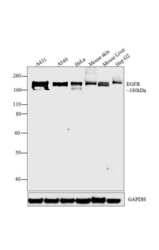
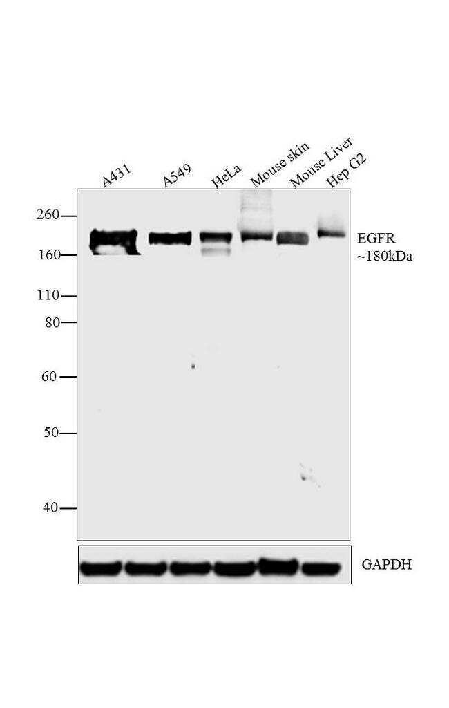
- Western blot analysis was performed on whole cell extracts (30 µg lysate) of A431 (Lane 1), A549 (Lane 2), HeLa (lane 3), Mouse Skin (Lane 4), Mouse Liver (Lane 5) and Hep G2 (Lane 6). The blots were probed with Phospho-EGFR (Tyr1068) Rabbit Monoclonal Antibody (Product # MA5-14485, 1:250 dilution) and detected by chemiluminescence Goat anti-Rabbit IgG (H+L) Secondary Antibody, HRP conjugate (Product # G-21234, 1:5,000 dilution). A 180 kDa band corresponding to EGFR was observed across cell lines and tissues tested. Known quantity of protein samples were electrophoresed using Novex® NuPAGE® 10 % Bis-Tris gel (Product # NP0301BOX), XCell SureLock™ Electrophoresis System (Product # EI0002) and Novex® Sharp Pre-Stained Protein Standard (Product # LC5800). Resolved proteins were then transferred onto a nitrocellulose membrane with by overnight wet transfer method. The membrane was probed with the relevant primary and secondary Antibody following blocking with 5 % skimmed milk. Chemiluminescent detection was performed using Pierce™ ECL Western Blotting Substrate (Product # 32106).
Supportive validation
- Submitted by
- Invitrogen Antibodies (provider)
- Main image
- Experimental details
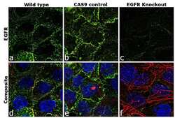
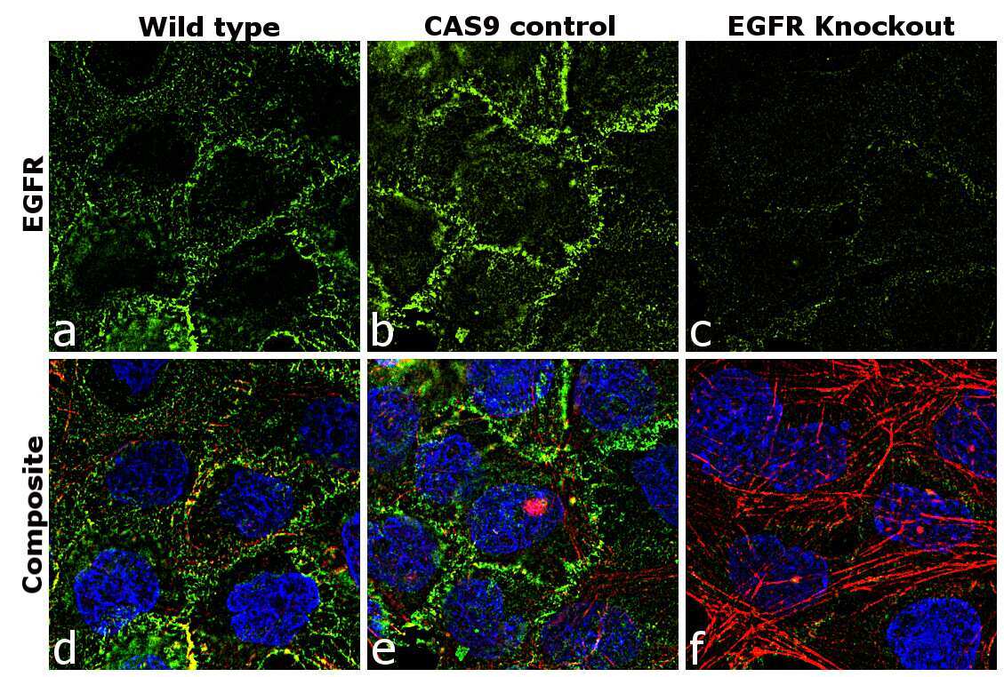
- Immunofluorescence analysis of EGFR was performed using 70% confluent log phase A-431 cells (WIld type, panels a,d), CAS9 control (panels b,e) and EGFR Knockout (panels c,f). The cells were fixed, permeabilized, and labelled with Phospho-EGFR (Tyr1068) Rabbit Monoclonal Antibody(Product # MA5-14485, 1:100), followed by Goat anti-Rabbit IgG (H+L) Superclonal™ Secondary Antibody, Alexa Fluor® 488 conjugate (Product # A27034, 1:2,000). Nuclei (blue) were stained with SlowFade® Gold Antifade Mountant with DAPI (Product # S36938) and Rhodamine Phalloidin (Product # R415, 1:300) was used for cytoskeletal F-actin (red) staining. Loss of signal was observed in EGFR Knockout cells (panel c,f) confirming specificity of the antibody to EGFR(green). The images were captured at 60X magnification.
- Submitted by
- Invitrogen Antibodies (provider)
- Main image
- Experimental details
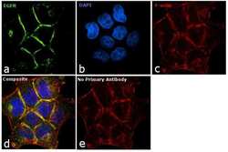
- Immunofluorescence analysis of EGFR was performed using 90% confluent log phase A431 cells. The cells were fixed with 4% paraformaldehyde for 10 minutes, permeabilized with 0.1% Triton™ X-100 for 10 minutes, and blocked with 1% BSA for 1 hour at room temperature. The cells were labeled with Phospho-EGFR (Tyr1068) Rabbit Monoclonal antibody (Product # MA5-14485) at 1:250 dilution in 0.1% BSA and incubated for 3 hours at room temperature and then labeled with Goat anti-Rabbit IgG (H+L) Superclonal™ Secondary Antibody, Alexa Fluor® 488 conjugate (Product # A27034) at a dilution of 1:2,000 for 45 minutes at room temperature (Panel a: green). Nuclei (Panel b: blue) were stained with SlowFade® Gold Antifade Mountant with DAPI (Product # S36938). F-actin (Panel c: red) was stained with Rhodamine Phalloidin (Product # R415, 1:300). Panel d represents the merged image showing membrane localization. Panel e shows the no primary antibody control. The images were captured at 60X magnification.

- Submitted by
- Invitrogen Antibodies (provider)
- Main image
- Experimental details
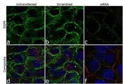
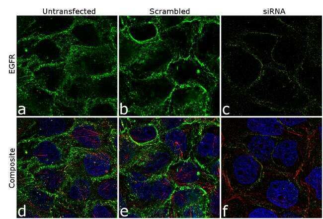
- Knockdown of EGFR was achieved by transfecting A431 cells with EGFR specific siRNA (Silencer® select Product # s563, s564 and s565). Immunofluorescence analysis was performed on A431 cells (untransfected, panel a,d), transfected with non-specific scrambled siRNA (panels b,e) and transfected with EGFR specific siRNA (panel c,f) Cells were fixed, permeabilized, and labelled with Phospho-EGFR (Tyr1068) Rabbit Monoclonal Antibody (Product # MA5-14485, 5 µg/mL), followed by Goat anti-Rabbit IgG (H+L) Superclonal™ Secondary Antibody, Alexa Fluor® 488 conjugate (Product # A27034, 1:2,000). Nuclei (blue) were stained using SlowFade® Gold Antifade Mountant with DAPI (Product # S36938), and Rhodamine Phalloidin (Product # R415, 1:300) was used for cytoskeletal F-actin (red) staining. Loss of signal was observed upon siRNA mediated knockdown (panel c,f) confirming specificity of the antibody to EGFR (green). The images were captured at 60X magnification.
Supportive validation
- Submitted by
- Invitrogen Antibodies (provider)
- Main image
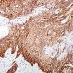
- Experimental details
- Formalin-fixed, paraffin-embedded human squamous cell carcinoma stained with rabbit monoclonal Phospho-EGFR (Tyr1068) antibody using peroxidase-conjugate and DAB chromogen. Note membrane staining of tumor cells.

Supportive validation
- Submitted by
- Invitrogen Antibodies (provider)
- Main image
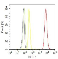
- Experimental details
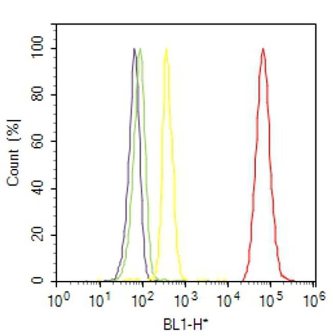
- Flow cytometry analysis of EGFR was done on A-431 cells. Cells were fixed with 70% ethanol for 10 minutes, permeabilized with 0.25% Triton™ X-100 for 20 minutes, and blocked with 5% BSA for 30 minutes at room temperature. Cells were labeled with EGFR Rabbit Monoclonal Antibody (MA514485, red histogram) or with rabbit isotype control (yellow histogram) at 3-5 ug/million cells in 2.5% BSA. After incubation at room temperature for 2 hours, the cells were labeled with Alexa Fluor® 488 Goat Anti-Rabbit Secondary Antibody (A11008) at a dilution of 1:400 for 30 minutes at room temperature. The representative 10,000 cells were acquired and analyzed for each sample using an Attune® Acoustic Focusing Cytometer. The purple histogram represents unstained control cells and the green histogram represents no-primary-antibody control.