Explore
 Validate
 Learn
Learn Immunocytochemistry
Antibody data
- Antibody Data
- Antigen structure
- References [28]
- Comments [0]
- Validations
- Immunocytochemistry [12]
- Immunoprecipitation [1]
- Other assay [2]
Submit
Validation data
Reference
Comment
Report error
- Product number
- MA5-12880 - Provider product page

- Provider
- Invitrogen Antibodies
- Product name
- EGFR Monoclonal Antibody (225)
- Antibody type
- Monoclonal
- Antigen
- Purifed from natural sources
- Description
- MA5-12880 targets Epidermal Growth Factor Receptor in IP and IF applications and shows reactivity with Human and Non-human primate samples. The MA5-12880 immunogen is purified EGFR from A431 cells.
- Reactivity
- Human
- Host
- Mouse
- Isotype
- IgG
- Antibody clone number
- 225
- Vial size
- 500 μL
- Concentration
- 0.2 mg/mL
- Storage
- 4°C
Submitted references Litmus-Body: A Molecularly Targeted Sensor for Cell-Surface pH Measurements.
IKKα regulates human keratinocyte migration through surveillance of the redox environment.
Sensitization of EGFR Wild-Type Non-Small Cell Lung Cancer Cells to EGFR-Tyrosine Kinase Inhibitor Erlotinib.
Interaction of EGFR to δ-catenin leads to δ-catenin phosphorylation and enhances EGFR signaling.
PHD4 stimulates tumor angiogenesis in osteosarcoma cells via TGF-α.
In vitro perforation of human epithelial carcinoma cell with antibody-conjugated biodegradable microspheres illuminated by a single 80 femtosecond near-infrared laser pulse.
Epidermal growth factor receptor as a biomarker for cervical cancer.
The β-catenin/Tcf4/survivin signaling maintains a less differentiated phenotype and high proliferative capacity of human corneal epithelial progenitor cells.
Receptor heterodimerization: a new mechanism for platelet-derived growth factor induced resistance to anti-epidermal growth factor receptor therapy for bladder cancer.
An EGFR autocrine loop encodes a slow-reacting but dominant mode of mechanotransduction in a polarized epithelium.
The antimicrobial peptide LL37 induces the migration of human pulp cells: a possible adjunct for regenerative endodontics.
Nano-bio-chip sensor platform for examination of oral exfoliative cytology.
Diminished survival of human cytotrophoblast cells exposed to hypoxia/reoxygenation injury and associated reduction of heparin-binding epidermal growth factor-like growth factor.
Involvement of membrane-type bile acid receptor M-BAR/TGR5 in bile acid-induced activation of epidermal growth factor receptor and mitogen-activated protein kinases in gastric carcinoma cells.
Epidermal growth factor-like growth factors prevent apoptosis of alcohol-exposed human placental cytotrophoblast cells.
Receptor-selective retinoids inhibit the growth of normal and malignant breast cells by inducing G1 cell cycle blockade.
Directed evolution of the epidermal growth factor receptor extracellular domain for expression in yeast.
Human trophoblast survival at low oxygen concentrations requires metalloproteinase-mediated shedding of heparin-binding EGF-like growth factor.
Inhibition of xenograft tumor growth and down-regulation of ErbB receptors by an antibody directed against Lewis Y antigen.
The role of LIP5 and CHMP5 in multivesicular body formation and HIV-1 budding in mammalian cells.
Autocrine extracellular signal-regulated kinase (ERK) activation in normal human keratinocytes: metalloproteinase-mediated release of amphiregulin triggers signaling from ErbB1 to ERK.
Fine epitope mapping of anti-epidermal growth factor receptor antibodies through random mutagenesis and yeast surface display.
Domain-level antibody epitope mapping through yeast surface display of epidermal growth factor receptor fragments.
Broadly distributed chemical reactivity of natural antibodies expressed in coordination with specific antigen binding activity.
Autoantibodies to the epidermal growth factor receptor in systemic sclerosis, lupus, and autoimmune mice.
Heparin-binding epidermal growth factor-like growth factor: hypoxia-inducible expression in vitro and stimulation of neurogenesis in vitro and in vivo.
Interaction between tyrosine kinase Etk and a RUN domain- and FYVE domain-containing protein RUFY1. A possible role of ETK in regulation of vesicle trafficking.
Differential utilization and localization of ErbB receptor tyrosine kinases in skin compared to normal and malignant keratinocytes.
Kuo JC, Goudge MC, Metzloff AE, Huang LT, Colville MJ, Park S, Zipfel WR, Paszek MJ
ACS sensors 2020 Jun 26;5(6):1555-1566
ACS sensors 2020 Jun 26;5(6):1555-1566
IKKα regulates human keratinocyte migration through surveillance of the redox environment.
Lisse TS, Rieger S
Journal of cell science 2017 Mar 1;130(5):975-988
Journal of cell science 2017 Mar 1;130(5):975-988
Sensitization of EGFR Wild-Type Non-Small Cell Lung Cancer Cells to EGFR-Tyrosine Kinase Inhibitor Erlotinib.
Raimbourg J, Joalland MP, Cabart M, de Plater L, Bouquet F, Savina A, Decaudin D, Bennouna J, Vallette FM, Lalier L
Molecular cancer therapeutics 2017 Aug;16(8):1634-1644
Molecular cancer therapeutics 2017 Aug;16(8):1634-1644
Interaction of EGFR to δ-catenin leads to δ-catenin phosphorylation and enhances EGFR signaling.
He Y, Ryu T, Shrestha N, Yuan T, Kim H, Shrestha H, Cho YC, Seo YW, Song WK, Kim K
Scientific reports 2016 Feb 17;6:21207
Scientific reports 2016 Feb 17;6:21207
PHD4 stimulates tumor angiogenesis in osteosarcoma cells via TGF-α.
Klotzsche-von Ameln A, Prade I, Grosser M, Kettelhake A, Rezaei M, Chavakis T, Flamme I, Wielockx B, Breier G
Molecular cancer research : MCR 2013 Nov;11(11):1337-48
Molecular cancer research : MCR 2013 Nov;11(11):1337-48
In vitro perforation of human epithelial carcinoma cell with antibody-conjugated biodegradable microspheres illuminated by a single 80 femtosecond near-infrared laser pulse.
Terakawa M, Tsunoi Y, Mitsuhashi T
International journal of nanomedicine 2012;7:2653-60
International journal of nanomedicine 2012;7:2653-60
Epidermal growth factor receptor as a biomarker for cervical cancer.
Soonthornthum T, Arias-Pulido H, Joste N, Lomo L, Muller C, Rutledge T, Verschraegen C
Annals of oncology : official journal of the European Society for Medical Oncology 2011 Oct;22(10):2166-78
Annals of oncology : official journal of the European Society for Medical Oncology 2011 Oct;22(10):2166-78
The β-catenin/Tcf4/survivin signaling maintains a less differentiated phenotype and high proliferative capacity of human corneal epithelial progenitor cells.
Lu R, Bian F, Zhang X, Qi H, Chuang EY, Pflugfelder SC, Li DQ
The international journal of biochemistry & cell biology 2011 May;43(5):751-9
The international journal of biochemistry & cell biology 2011 May;43(5):751-9
Receptor heterodimerization: a new mechanism for platelet-derived growth factor induced resistance to anti-epidermal growth factor receptor therapy for bladder cancer.
Black PC, Brown GA, Dinney CP, Kassouf W, Inamoto T, Arora A, Gallagher D, Munsell MF, Bar-Eli M, McConkey DJ, Adam L
The Journal of urology 2011 Feb;185(2):693-700
The Journal of urology 2011 Feb;185(2):693-700
An EGFR autocrine loop encodes a slow-reacting but dominant mode of mechanotransduction in a polarized epithelium.
Kojic N, Chung E, Kho AT, Park JA, Huang A, So PT, Tschumperlin DJ
FASEB journal : official publication of the Federation of American Societies for Experimental Biology 2010 May;24(5):1604-15
FASEB journal : official publication of the Federation of American Societies for Experimental Biology 2010 May;24(5):1604-15
The antimicrobial peptide LL37 induces the migration of human pulp cells: a possible adjunct for regenerative endodontics.
Kajiya M, Shiba H, Komatsuzawa H, Ouhara K, Fujita T, Takeda K, Uchida Y, Mizuno N, Kawaguchi H, Kurihara H
Journal of endodontics 2010 Jun;36(6):1009-13
Journal of endodontics 2010 Jun;36(6):1009-13
Nano-bio-chip sensor platform for examination of oral exfoliative cytology.
Weigum SE, Floriano PN, Redding SW, Yeh CK, Westbrook SD, McGuff HS, Lin A, Miller FR, Villarreal F, Rowan SD, Vigneswaran N, Williams MD, McDevitt JT
Cancer prevention research (Philadelphia, Pa.) 2010 Apr;3(4):518-28
Cancer prevention research (Philadelphia, Pa.) 2010 Apr;3(4):518-28
Diminished survival of human cytotrophoblast cells exposed to hypoxia/reoxygenation injury and associated reduction of heparin-binding epidermal growth factor-like growth factor.
Leach RE, Kilburn BA, Petkova A, Romero R, Armant DR
American journal of obstetrics and gynecology 2008 Apr;198(4):471.e1-7; discussion 471.e7-8
American journal of obstetrics and gynecology 2008 Apr;198(4):471.e1-7; discussion 471.e7-8
Involvement of membrane-type bile acid receptor M-BAR/TGR5 in bile acid-induced activation of epidermal growth factor receptor and mitogen-activated protein kinases in gastric carcinoma cells.
Yasuda H, Hirata S, Inoue K, Mashima H, Ohnishi H, Yoshiba M
Biochemical and biophysical research communications 2007 Mar 2;354(1):154-9
Biochemical and biophysical research communications 2007 Mar 2;354(1):154-9
Epidermal growth factor-like growth factors prevent apoptosis of alcohol-exposed human placental cytotrophoblast cells.
Wolff GS, Chiang PJ, Smith SM, Romero R, Armant DR
Biology of reproduction 2007 Jul;77(1):53-60
Biology of reproduction 2007 Jul;77(1):53-60
Receptor-selective retinoids inhibit the growth of normal and malignant breast cells by inducing G1 cell cycle blockade.
Wu K, DuPré E, Kim H, Tin-U CK, Bissonnette RP, Lamph WW, Brown PH
Breast cancer research and treatment 2006 Mar;96(2):147-57
Breast cancer research and treatment 2006 Mar;96(2):147-57
Directed evolution of the epidermal growth factor receptor extracellular domain for expression in yeast.
Kim YS, Bhandari R, Cochran JR, Kuriyan J, Wittrup KD
Proteins 2006 Mar 1;62(4):1026-35
Proteins 2006 Mar 1;62(4):1026-35
Human trophoblast survival at low oxygen concentrations requires metalloproteinase-mediated shedding of heparin-binding EGF-like growth factor.
Armant DR, Kilburn BA, Petkova A, Edwin SS, Duniec-Dmuchowski ZM, Edwards HJ, Romero R, Leach RE
Development (Cambridge, England) 2006 Feb;133(4):751-9
Development (Cambridge, England) 2006 Feb;133(4):751-9
Inhibition of xenograft tumor growth and down-regulation of ErbB receptors by an antibody directed against Lewis Y antigen.
Farhan H, Schuster C, Klinger M, Weisz E, Waxenecker G, Schuster M, Sexl V, Mudde GC, Freissmuth M, Kircheis R
The Journal of pharmacology and experimental therapeutics 2006 Dec;319(3):1459-66
The Journal of pharmacology and experimental therapeutics 2006 Dec;319(3):1459-66
The role of LIP5 and CHMP5 in multivesicular body formation and HIV-1 budding in mammalian cells.
Ward DM, Vaughn MB, Shiflett SL, White PL, Pollock AL, Hill J, Schnegelberger R, Sundquist WI, Kaplan J
The Journal of biological chemistry 2005 Mar 18;280(11):10548-55
The Journal of biological chemistry 2005 Mar 18;280(11):10548-55
Autocrine extracellular signal-regulated kinase (ERK) activation in normal human keratinocytes: metalloproteinase-mediated release of amphiregulin triggers signaling from ErbB1 to ERK.
Kansra S, Stoll SW, Johnson JL, Elder JT
Molecular biology of the cell 2004 Sep;15(9):4299-309
Molecular biology of the cell 2004 Sep;15(9):4299-309
Fine epitope mapping of anti-epidermal growth factor receptor antibodies through random mutagenesis and yeast surface display.
Chao G, Cochran JR, Wittrup KD
Journal of molecular biology 2004 Sep 10;342(2):539-50
Journal of molecular biology 2004 Sep 10;342(2):539-50
Domain-level antibody epitope mapping through yeast surface display of epidermal growth factor receptor fragments.
Cochran JR, Kim YS, Olsen MJ, Bhandari R, Wittrup KD
Journal of immunological methods 2004 Apr;287(1-2):147-58
Journal of immunological methods 2004 Apr;287(1-2):147-58
Broadly distributed chemical reactivity of natural antibodies expressed in coordination with specific antigen binding activity.
Planque S, Taguchi H, Burr G, Bhatia G, Karle S, Zhou YX, Nishiyama Y, Paul S
The Journal of biological chemistry 2003 May 30;278(22):20436-43
The Journal of biological chemistry 2003 May 30;278(22):20436-43
Autoantibodies to the epidermal growth factor receptor in systemic sclerosis, lupus, and autoimmune mice.
Planque S, Zhou YX, Nishiyama Y, Sinha M, O'Connor-Mccourt M, Arnett FC, Paul S
FASEB journal : official publication of the Federation of American Societies for Experimental Biology 2003 Feb;17(2):136-43
FASEB journal : official publication of the Federation of American Societies for Experimental Biology 2003 Feb;17(2):136-43
Heparin-binding epidermal growth factor-like growth factor: hypoxia-inducible expression in vitro and stimulation of neurogenesis in vitro and in vivo.
Jin K, Mao XO, Sun Y, Xie L, Jin L, Nishi E, Klagsbrun M, Greenberg DA
The Journal of neuroscience : the official journal of the Society for Neuroscience 2002 Jul 1;22(13):5365-73
The Journal of neuroscience : the official journal of the Society for Neuroscience 2002 Jul 1;22(13):5365-73
Interaction between tyrosine kinase Etk and a RUN domain- and FYVE domain-containing protein RUFY1. A possible role of ETK in regulation of vesicle trafficking.
Yang J, Kim O, Wu J, Qiu Y
The Journal of biological chemistry 2002 Aug 16;277(33):30219-26
The Journal of biological chemistry 2002 Aug 16;277(33):30219-26
Differential utilization and localization of ErbB receptor tyrosine kinases in skin compared to normal and malignant keratinocytes.
Stoll SW, Kansra S, Peshick S, Fry DW, Leopold WR, Wiesen JF, Sibilia M, Zhang T, Werb Z, Derynck R, Wagner EF, Elder JT
Neoplasia (New York, N.Y.) 2001 Jul-Aug;3(4):339-50
Neoplasia (New York, N.Y.) 2001 Jul-Aug;3(4):339-50
No comments: Submit comment
Supportive validation
- Submitted by
- Invitrogen Antibodies (provider)
- Main image

- Experimental details
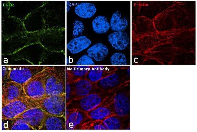
- Immunofluorescence analysis of EGFR was performed using 90% confluent log phase A431 cells. The cells were fixed with 4% paraformaldehyde for 10 minutes, permeabilized with 0.1% Triton™ X-100 for 10 minutes, and blocked with 1% BSA for 1 hour at room temperature. The cells were labeled with EGFR Mouse monoclonal antibody (Product # MA5-12880) at 5 µg/mL in 0.1% BSA and incubated for 3 hours at room temperature and then labeled with Goat anti-Mouse IgG (H+L) Superclonal™ Secondary Antibody, Alexa Fluor® 488 conjugate (Product # A28175) at a dilution of 1:2000 for 45 minutes at room temperature (Panel a: green). Nuclei (Panel b: blue) were stained with SlowFade® Gold Antifade Mountant with DAPI (Product # S36938). F-actin (Panel c: red) was stained with Rhodamine Phalloidin (Product # R415, 1:300). Panel d represents the merged image showing membrane localization. Panel e shows the no primary antibody control. The images were captured at 60X magnification.
- Submitted by
- Invitrogen Antibodies (provider)
- Main image
- Experimental details
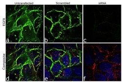
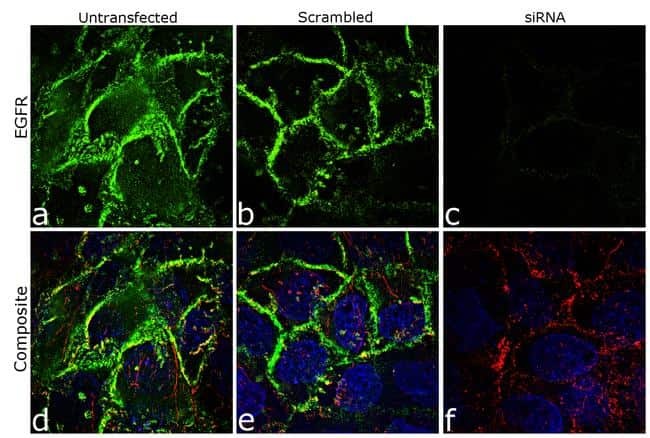
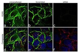
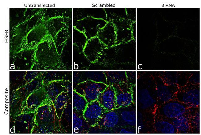
- Knockdown of EGFR was achieved by transfecting A-431 cells with EGFR specific siRNA (Silencer® select Cat # s563, s564, s565). Immunofluorescence analysis was performed on A431 cells (untransfected, panel a,d), transfected with non-specific scrambled siRNA (panels b,e) and transfected with EGFR specific siRNA (panel c,f) Cells were fixed, permeabilized, and labelled with EGFR Mouse monoclonal Antibody (Product # MA5-12880, 5 µg/mL), followed by Goat anti-Mouse IgG (H+L) Superclonal™ Secondary Antibody, Alexa Fluor® 488 conjugate (Product # A28175, 1:2000). Nuclei (blue) were stained using SlowFade® Gold Antifade Mountant with DAPI (Product # S36938), and Rhodamine Phalloidin (Product # R415, 1:300) was used for cytoskeletal F-actin (red) staining. Loss of signal was observed upon siRNA mediated knockdown (panel c,f) confirming specificity of the antibody to EGFR(green). The images were captured at 60X magnification.
- Submitted by
- Invitrogen Antibodies (provider)
- Main image
- Experimental details
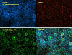
- Immunofluorescent analysis of Phalloidin (blue) and EGFR (green) in A431 cells. Formalin fixed cells were permeabilized with 0.1% Triton X-100 in PBS for 10 minutes at room temperature and blocked with 2% BSA in PBS + 0.1% Triton X-100 (Product # 37525) for 30 minutes at room temperature. Cells were probed with an EGFR monoclonal antibody (Product # MA5-12880) at a dilution of 1:75 for at least 1 hour at room temperature, washed with PBS, and incubated with Dylight 488 goat anti-mouse IgG secondary antibody (Product # 35502) at a dilution of 1:250 for 30 minutes at room temperature. Actin was stained with Dylight 350 Phalloidin (Product # 21830) at a dilution of 1:120 (2.5units/mL final concentration) and nuclei (red) were stained with DRAQ5 (Product # 62254) at a concentration of 1 µg/mL for 30 minutes. Images were taken on a Zeiss Axio Observer Z1 microscope at 20X magnification.
- Submitted by
- Invitrogen Antibodies (provider)
- Main image

- Experimental details
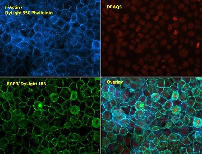
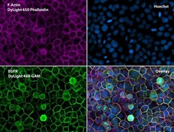
- Immunofluorescent analysis of Phalloidin (orange) and EGFR (green) in A431 cells. Formalin fixed cells were permeabilized with 0.1% Triton X-100 in PBS for 10 minutes at room temperature and blocked with 2% BSA (Product # 37525) in PBS + 0.1% Triton X-100 for 30 minutes at room temperature. Cells were probed with an EGFR monoclonal antibody (Product # MA5-12880) at a dilution of 1:75 for at least 1 hour at room temperature, washed with PBS, and incubated with DyLight 488 goat anti-mouse IgG secondary antibody (Product # 35502) at a dilution of 1:250 for 30 minutes at room temperature. Actin was stained with DyLight 550 Phalloidin (Product # 21835) at a dilution of 1:120 (2.5 units/mL final concentration) and nuclei (blue) were stained with Hoechst (Product # 62249) at a concentration of 1 µg/mL for 30 minutes. Images were taken on a Zeiss Axio Observer Z1 microscope at 20X magnification.

- Submitted by
- Invitrogen Antibodies (provider)
- Main image

- Experimental details
- Immunofluorescent analysis of Phalloidin (purple) and EGFR (green) in A431 cells. Formalin fixed cells were permeabilized with 0.1% Triton X-100 in PBS for 10 minutes at room temperature and blocked with 2% BSA (Product # 37525) in PBS + 0.1% Triton X-100 for 30 minutes at room temperature. Cells were probed with an EGFR monoclonal antibody (Product # MA5-12880) at a dilution of 1:75 for at least 1 hour at room temperature, washed with PBS, and incubated with DyLight 488 goat anti-mouse IgG secondary antibody (Product # 35502) at a dilution of 1:250 for 30 minutes at room temperature. Actin was stained with DyLight 650 Phalloidin (Product # 21838) at a dilution of 1:120 (2.5 units/mL final concentration) and nuclei (blue) were stained with Hoechst (Product # 62249) at a concentration of 1 µg/mL for 30 minutes. Images were taken on a Zeiss Axio Observer Z1 microscope at 20X magnification.
- Submitted by
- Invitrogen Antibodies (provider)
- Main image
- Experimental details
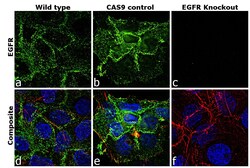
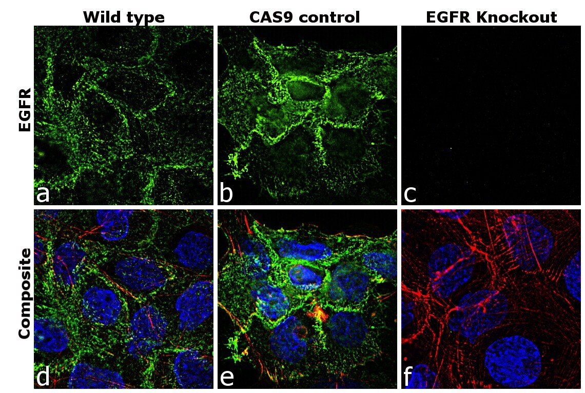
- Immunofluorescence analysis of EGFR was performed using 70% confluent log phase A-431 cells (WIld type, panels a,d), CAS9 control (panels b,e) and EGFR Knockout (panels c,f). The cells were fixed, permeabilized, and labelled with EGFR Mouse Monoclonal Antibody(Product # MA5-12880, 5 µg/mL), followed by Goat anti-Mouse IgG (H+L) Superclonal™ Secondary Antibody, Alexa Fluor® 488 conjugate (Product # A28175, 1:2000). Nuclei (blue) were stained with SlowFade® Gold Antifade Mountant with DAPI (Product # S36938) and Rhodamine Phalloidin (Product # R415, 1:300) was used for cytoskeletal F-actin (red) staining. Loss of signal was observed in EGFR Knockout cells (panel c,f) confirming specificity of the antibody to EGFR(green). The images were captured at 60X magnification.

- Submitted by
- Invitrogen Antibodies (provider)
- Main image

- Experimental details
- Immunofluorescent analysis of Phalloidin (blue) and EGFR (green) in A431 cells. Formalin fixed cells were permeabilized with 0.1% Triton X-100 in PBS for 10 minutes at room temperature and blocked with 2% BSA in PBS + 0.1% Triton X-100 (Product # 37525) for 30 minutes at room temperature. Cells were probed with an EGFR monoclonal antibody (Product # MA5-12880) at a dilution of 1:75 for at least 1 hour at room temperature, washed with PBS, and incubated with Dylight 488 goat anti-mouse IgG secondary antibody (Product # 35502) at a dilution of 1:250 for 30 minutes at room temperature. Actin was stained with Dylight 350 Phalloidin (Product # 21830) at a dilution of 1:120 (2.5units/mL final concentration) and nuclei (red) were stained with DRAQ5 (Product # 62254) at a concentration of 1 µg/mL for 30 minutes. Images were taken on a Zeiss Axio Observer Z1 microscope at 20X magnification.

- Submitted by
- Invitrogen Antibodies (provider)
- Main image

- Experimental details
- Immunofluorescent analysis of Phalloidin (orange) and EGFR (green) in A431 cells. Formalin fixed cells were permeabilized with 0.1% Triton X-100 in PBS for 10 minutes at room temperature and blocked with 2% BSA (Product # 37525) in PBS + 0.1% Triton X-100 for 30 minutes at room temperature. Cells were probed with an EGFR monoclonal antibody (Product # MA5-12880) at a dilution of 1:75 for at least 1 hour at room temperature, washed with PBS, and incubated with DyLight 488 goat anti-mouse IgG secondary antibody (Product # 35502) at a dilution of 1:250 for 30 minutes at room temperature. Actin was stained with DyLight 550 Phalloidin (Product # 21835) at a dilution of 1:120 (2.5 units/mL final concentration) and nuclei (blue) were stained with Hoechst (Product # 62249) at a concentration of 1 µg/mL for 30 minutes. Images were taken on a Zeiss Axio Observer Z1 microscope at 20X magnification.

- Submitted by
- Invitrogen Antibodies (provider)
- Main image
- Experimental details
- Immunofluorescent analysis of Phalloidin (purple) and EGFR (green) in A431 cells. Formalin fixed cells were permeabilized with 0.1% Triton X-100 in PBS for 10 minutes at room temperature and blocked with 2% BSA (Product # 37525) in PBS + 0.1% Triton X-100 for 30 minutes at room temperature. Cells were probed with an EGFR monoclonal antibody (Product # MA5-12880) at a dilution of 1:75 for at least 1 hour at room temperature, washed with PBS, and incubated with DyLight 488 goat anti-mouse IgG secondary antibody (Product # 35502) at a dilution of 1:250 for 30 minutes at room temperature. Actin was stained with DyLight 650 Phalloidin (Product # 21838) at a dilution of 1:120 (2.5 units/mL final concentration) and nuclei (blue) were stained with Hoechst (Product # 62249) at a concentration of 1 µg/mL for 30 minutes. Images were taken on a Zeiss Axio Observer Z1 microscope at 20X magnification.

- Submitted by
- Invitrogen Antibodies (provider)
- Main image
- Experimental details
- Knockdown of EGFR was achieved by transfecting A-431 cells with EGFR specific siRNA (Silencer® select Cat # s563, s564, s565). Immunofluorescence analysis was performed on A431 cells (untransfected, panel a,d), transfected with non-specific scrambled siRNA (panels b,e) and transfected with EGFR specific siRNA (panel c,f) Cells were fixed, permeabilized, and labelled with EGFR Mouse monoclonal Antibody (Product # MA5-12880, 5 µg/mL), followed by Goat anti-Mouse IgG (H+L) Superclonal™ Secondary Antibody, Alexa Fluor® 488 conjugate (Product # A28175, 1:2000). Nuclei (blue) were stained using SlowFade® Gold Antifade Mountant with DAPI (Product # S36938), and Rhodamine Phalloidin (Product # R415, 1:300) was used for cytoskeletal F-actin (red) staining. Loss of signal was observed upon siRNA mediated knockdown (panel c,f) confirming specificity of the antibody to EGFR(green). The images were captured at 60X magnification.
- Submitted by
- Invitrogen Antibodies (provider)
- Main image

- Experimental details
- Immunofluorescence analysis of EGFR was performed using 90% confluent log phase A431 cells. The cells were fixed with 4% paraformaldehyde for 10 minutes, permeabilized with 0.1% Triton™ X-100 for 10 minutes, and blocked with 1% BSA for 1 hour at room temperature. The cells were labeled with EGFR Mouse monoclonal antibody (Product # MA5-12880) at 5 µg/mL in 0.1% BSA and incubated for 3 hours at room temperature and then labeled with Goat anti-Mouse IgG (H+L) Superclonal™ Secondary Antibody, Alexa Fluor® 488 conjugate (Product # A28175) at a dilution of 1:2000 for 45 minutes at room temperature (Panel a: green). Nuclei (Panel b: blue) were stained with SlowFade® Gold Antifade Mountant with DAPI (Product # S36938). F-actin (Panel c: red) was stained with Rhodamine Phalloidin (Product # R415, 1:300). Panel d represents the merged image showing membrane localization. Panel e shows the no primary antibody control. The images were captured at 60X magnification.

- Submitted by
- Invitrogen Antibodies (provider)
- Main image

- Experimental details
- Immunofluorescence analysis of EGFR was performed using 70% confluent log phase A-431 cells (WIld type, panels a,d), CAS9 control (panels b,e) and EGFR Knockout (panels c,f). The cells were fixed, permeabilized, and labelled with EGFR Mouse Monoclonal Antibody(Product # MA5-12880, 5 µg/mL), followed by Goat anti-Mouse IgG (H+L) Superclonal™ Secondary Antibody, Alexa Fluor® 488 conjugate (Product # A28175, 1:2000). Nuclei (blue) were stained with SlowFade® Gold Antifade Mountant with DAPI (Product # S36938) and Rhodamine Phalloidin (Product # R415, 1:300) was used for cytoskeletal F-actin (red) staining. Loss of signal was observed in EGFR Knockout cells (panel c,f) confirming specificity of the antibody to EGFR(green). The images were captured at 60X magnification.
Supportive validation
- Submitted by
- Invitrogen Antibodies (provider)
- Main image
- Experimental details
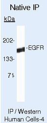
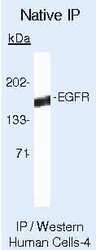
- Immunoprecipitation of Epidermal Growth Factor Receptor using Epidermal Growth Factor Receptor Monoclonal Antibody (Product # MA5-12880) on Native Human A431 Cells.
Supportive validation
- Submitted by
- Invitrogen Antibodies (provider)
- Main image
- Experimental details
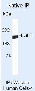
- Immunoprecipitation of Epidermal Growth Factor Receptor using Epidermal Growth Factor Receptor Monoclonal Antibody (Product # MA5-12880) on Native Human A431 Cells.

- Submitted by
- Invitrogen Antibodies (provider)
- Main image
- Experimental details
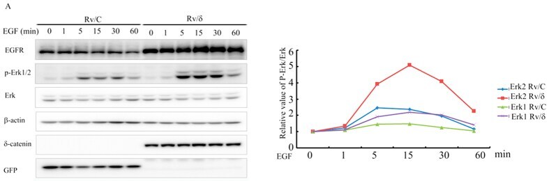
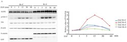
- Figure 8 delta-Catenin enhanced the EGFR/Erk1/2 signaling in CWR22Rv-1 cells. Rv/C and Rv/delta cells were cultured in serum free condition for 16 h followed by treatment of 100 nM EGF for different duration, as indicated. The cells were harvested and subjected to immunoblotting with the following antibodies: EGFR, p-Erk1/2, Erk1 (detect both Erk1 and Erk2), beta-actin, delta-catenin and GFP. These proteins were all from the same lysate. EGFR and beta-actin were from the same gel; GFP and delta-catenin were from the same gel; Erk1/2 and p-Erk1/2 were from the same gel. The experiments were performed 5 times. The density of p-Erk1/2 bands were normalized by the density of total Erk1/2 bands. The relative values were shown in the right panel.