 Explore
 Validate
 Learn
Learn Western blot
 ELISA
ELISAAntibody data
- Antibody Data
- Antigen structure
- References [0]
- Comments [0]
- Validations
- Western blot [1]
- Immunohistochemistry [1]
Submit
Validation data
Reference
Comment
Report error
- Product number
- AP09320PU-N - Provider product page

- Provider
- Acris Antibodies GmbH
- Proper citation
- Acris Antibodies GmbH Cat#AP09320PU-N, RRID:AB_2035448
- Product name
- anti EGFR / ERBB1 pTyr1197
- Antibody type
- Polyclonal
- Antigen
- Synthetic peptide corresponding to amino acids 1189-1199 of human EGFR protein
- Reactivity
- Human, Mouse, Rat
- Host
- Rabbit
- Isotype
- IgG
- Vial size
- 0.1 mg
- Concentration
- 0.55 mg/ml (by UV absorbance at 280 nm)
No comments: Submit comment
Supportive validation
- Submitted by
- Acris Antibodies GmbH (provider)
- Main image
- Experimental details
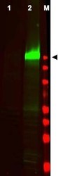
- Western blot using Affinity Purified anti-EGFR pY1197 antibody shows detection of a band at ~170 kDa corresponding to human EGFR (arrowhead). Staining is not seen in unstimulated A431 cells (lane 1), but is seen when A431 cells are stimulated with EGF (50 ng/ml for 15 min) (lane 2). Approximately 30 µg of lysate was separated on a 4-20% Tris-Glycine gel by SDS-PAGE and transferred onto nitrocellulose. After blocking the membrane was probed with the primary antibody diluted to 1:250. Reaction occurred overnight at 4° C followed by washes and reaction with a 1:10,000 dilution of IRDye(TM)800 conjugated Gt-a-Rabbit IgG [H&L] MX for 45 min at room temperature (800 nm channel, green). Molecular weight estimation was made by comparison to prestained MW markers in lane M (700 nm channel, red). IRDye(TM)800 fluorescence image was captured using the Odyssey(R) Infrared Imaging System developed by LI-COR. IRDye is a trademark of LI-COR, Inc. Other detection systems will yield similar results.

Supportive validation
- Submitted by
- Acris Antibodies GmbH (provider)
- Main image
- Experimental details
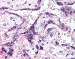
- Immunohistochemistry. Affinity purified anti-EGFR pY1197 antibody was used at 5 µg/ml to detect signal in a variety of tissues including multi-human, multi-brain and multicancer slides. This image shows faintly to moderately positive staining of placental trophoblasts at 40X. Tissue was formalinfixed and paraffin embedded. The image shows localization of the antibody as the precipitated red signal, with a hematoxylin purple nuclear counterstain.
