 Explore
 Validate
 Learn
Learn Western blot
 Immunocytochemistry
Antibody data
- Antibody Data
- Antigen structure
- References [1]
- Comments [0]
- Validations
- Western blot [3]
- Immunocytochemistry [1]
- Immunoprecipitation [1]
- Immunohistochemistry [2]
Submit
Validation data
Reference
Comment
Report error
- Product number
- GTX628887 - Provider product page

- Provider
- GeneTex
- Product name
- EGFR antibody [GT133]
- Antibody type
- Monoclonal
- Reactivity
- Human
- Host
- Mouse
Submitted references A high-throughput pipeline for validation of antibodies.
Sikorski K, Mehta A, Inngjerdingen M, Thakor F, Kling S, Kalina T, Nyman TA, Stensland ME, Zhou W, de Souza GA, Holden L, Stuchly J, Templin M, Lund-Johansen F
Nature methods 2018 Nov;15(11):909-912
Nature methods 2018 Nov;15(11):909-912
No comments: Submit comment
Enhanced validation
Supportive validation
- Submitted by
- GeneTex (provider)
- Enhanced method
- Genetic validation
- Main image
- Experimental details
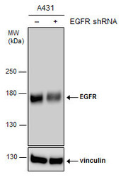
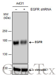
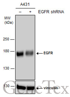
- Non-transfected (¡V) and transfected (+) A431 whole cell extracts (15 ?g) were separated by 5% SDS-PAGE, and the membrane was blotted with EGFR antibody [GT133] (GTX628887) diluted at 1:8000. The HRP-conjugated anti-mouse IgG antibody (GTX213111-01) was used to detect the primary antibody.
Supportive validation
- Submitted by
- GeneTex (provider)
- Main image
- Experimental details
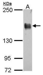
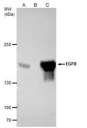
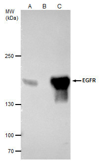
- EGFR antibody [GT133] detects EGFR protein by western blot analysis.A. 30 ?g A431 whole cell lysate/extract5% SDS-PAGEEGFR antibody [GT133] (GTX628887) dilution: 1:10000 The HRP-conjugated anti-mouse IgG antibody (GTX213111-01) was used to detect the primary antibody.

- Submitted by
- GeneTex (provider)
- Main image
- Experimental details
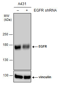
- Non-transfected (¡V) and transfected (+) A431 whole cell extracts (15 ?g) were separated by 5% SDS-PAGE, and the membrane was blotted with EGFR antibody [GT133] (GTX628887) diluted at 1:8000. The HRP-conjugated anti-mouse IgG antibody (GTX213111-01) was used to detect the primary antibody.
Supportive validation
- Submitted by
- GeneTex (provider)
- Main image
- Experimental details
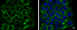
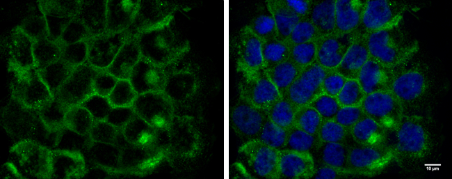
- EGFR antibody [GT133] detects EGFR protein at cell membrane by immunofluorescent analysis.Sample: A431 cells were fixed in 4% paraformaldehyde for 10 min.Green: EGFR protein stained by EGFR antibody [GT133] (GTX628887) diluted at 1:300.Blue: Hoechst 33342 staining.Scale bar = 10 £gm.
Supportive validation
- Submitted by
- GeneTex (provider)
- Main image
- Experimental details
- EGFR antibody [GT133] immunoprecipitates EGFR protein in IP experiments.IP samples: A431 whole cell extractA. 40 £gg A431 whole cell extractB. Control with 4 £gg of preimmune Mouse IgGC. Immunoprecipitation of EGFR protein by 4 £gg EGFR antibody [GT133] (GTX628887)5 % SDS-PAGEThe immunoprecipitated EGFR protein was detected by EGFR antibody [GT133] (GTX628887) diluted at 1:5000.[EasyBlot anti-mouse IgG (GTX221667-01) was used as a secondary reagent]
Supportive validation
- Submitted by
- GeneTex (provider)
- Main image
- Experimental details
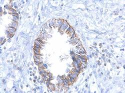
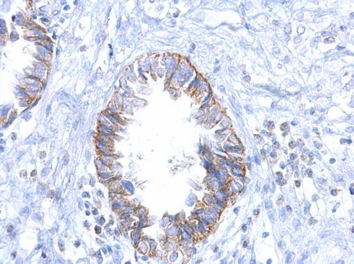
- EGFR antibody [GT133] detects EGFR protein at cytosol on human ovarian carcinoma by immunohistochemical analysis. Sample: Paraffin-embedded human ovarian carcinoma. EGFR antibody [GT133] (GTX628887) dilution: 1:500.
- Submitted by
- GeneTex (provider)
- Main image
- Experimental details
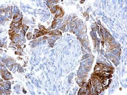
- EGFR antibody [GT133] detects EGFR protein at cytosol on human colon carcinoma by immunohistochemical analysis. Sample: Paraffin-embedded human colon carcinoma. EGFR antibody [GT133] (GTX628887) dilution: 1:500.
