 Explore
 Validate
 Learn
Learn Western blot
Antibody data
- Antibody Data
- Antigen structure
- References [2]
- Comments [0]
- Validations
- Western blot [2]
- Immunohistochemistry [1]
Submit
Validation data
Reference
Comment
Report error
- Product number
- AF5448 - Provider product page

- Provider
- Novus Biologicals
- Product name
- Sheep Polyclonal Syntaxin 8 Antibody
- Antibody type
- Polyclonal
- Description
- Immunogen affinity purified. Detects human, mouse, and rat Syntaxin 8 in Western blots.
- Reactivity
- Human, Mouse, Rat
- Host
- Sheep
- Isotype
- IgG
- Vial size
- 100 ug
- Concentration
- LYOPH
- Storage
- Use a manual defrost freezer and avoid repeated freeze-thaw cycles. 12 months from date of receipt, -20 to -70 degreesC as supplied. 1 month, 2 to 8 degreesC under sterile conditions after reconstitution. 6 months, -20 to -70 degreesC under sterile conditions after reconstitution.
Submitted references Munc13-4 functions as a Ca(2+) sensor for homotypic secretory granule fusion to generate endosomal exocytic vacuoles.
Syntaxin 8 regulates platelet dense granule secretion, aggregation, and thrombus stability.
Woo SS, James DJ, Martin TF
Molecular biology of the cell 2017 Mar 15;28(6):792-808
Molecular biology of the cell 2017 Mar 15;28(6):792-808
Syntaxin 8 regulates platelet dense granule secretion, aggregation, and thrombus stability.
Golebiewska EM, Harper MT, Williams CM, Savage JS, Goggs R, Fischer von Mollard G, Poole AW
The Journal of biological chemistry 2015 Jan 16;290(3):1536-45
The Journal of biological chemistry 2015 Jan 16;290(3):1536-45
No comments: Submit comment
Supportive validation
- Submitted by
- Novus Biologicals (provider)
- Main image
- Experimental details
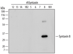
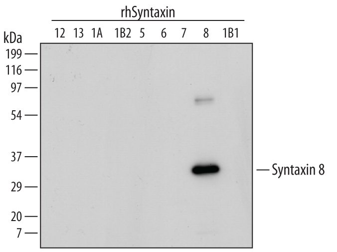
- Detection of Human Syntaxin 8 by Western Blot. Western blot shows recombinant human Syntaxin 12, 16, 1A, 1B2, 5, 6, 7, 8, and 1B1 (5 ng/lane). PVDF membrane was probed with 1 µg/mL Sheep Anti-Human/Mouse/Rat Syntaxin 8 Antigen Affinity-purified Polyclonal Antibody (Catalog # AF5448) followed by HRP-conjugated Anti-Sheep IgG Secondary Antibody (Catalog # HAF016). A specific band for Syntaxin 8 was detected as indicated. This experiment was conducted under reducing conditions and using Immunoblot Buffer Group 1.
- Submitted by
- Novus Biologicals (provider)
- Main image
- Experimental details
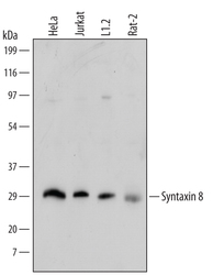
- Detection of Human, Mouse, and Rat Syntaxin 8 by Western Blot. Western blot shows lysates of HeLa human cervical epithelial carcinoma cell line, Jurkat human acute T cell leukemia cell line, L1.2 mouse pro-B cell line, and Rat-2 rat embryonic fibroblast cell line. PVDF membrane was probed with 1 µg/mL Sheep Anti-Human/Mouse/Rat Syntaxin 8 Antigen Affinity-purified Polyclonal Antibody (Catalog # AF5448) followed by HRP-conjugated Anti-Sheep IgG Secondary Antibody (Catalog # HAF016). A specific band for Syntaxin 8 was detected at approximately 28 kDa (as indicated). This experiment was conducted under reducing conditions and using Immunoblot Buffer Group 1.

Supportive validation
- Submitted by
- Novus Biologicals (provider)
- Main image
- Experimental details
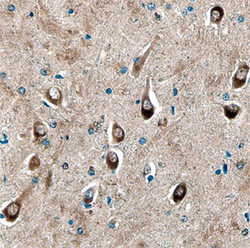
- Syntaxin 8 in Human Brain. Syntaxin 8 was detected in immersion fixed paraffin-embedded sections of human brain using Sheep Anti-Human/Mouse/Rat Syntaxin 8 Antigen Affinity-purified Polyclonal Antibody (Catalog # AF5448) at 10 µg/mL overnight at 4 °C. Before incubation with the primary antibody, tissue was subjected to heat-induced epitope retrieval using Antigen Retrieval Reagent-Basic (Catalog # CTS013). Tissue was stained using the Anti-Sheep HRP-DAB Cell & Tissue Staining Kit (brown; Catalog # CTS019) and counterstained with hematoxylin (blue). Specific staining was localized to neuronal cell bodies and processes. View our protocol for Chromogenic IHC Staining of Paraffin-embedded Tissue Sections.
