Explore
 Validate
 Learn
Learn Western blot
 Immunocytochemistry
Antibody data
- Antibody Data
- Antigen structure
- References [0]
- Comments [0]
- Validations
- Immunocytochemistry [4]
- Immunoprecipitation [1]
- Immunohistochemistry [3]
- Chromatin Immunoprecipitation [4]
- Other assay [1]
Submit
Validation data
Reference
Comment
Report error
- Product number
- PA5-28749 - Provider product page

- Provider
- Invitrogen Antibodies
- Product name
- ESRRA Polyclonal Antibody
- Antibody type
- Polyclonal
- Antigen
- Synthetic peptide
- Description
- Recommended positive controls: 293T, A431, H1299, Mouse kidney, Rat kidney, A549. Predicted reactivity: Mouse (100%), Rat (100%), Dog (100%), Pig (100%), Bovine (100%). Store product as a concentrated solution. Centrifuge briefly prior to opening the vial.
- Reactivity
- Human, Mouse, Rat
- Host
- Rabbit
- Isotype
- IgG
- Vial size
- 100 μL
- Concentration
- 1.32 mg/mL
- Storage
- Store at 4°C short term. For long term storage, store at -20°C, avoiding freeze/thaw cycles.
No comments: Submit comment
Supportive validation
- Submitted by
- Invitrogen Antibodies (provider)
- Main image

- Experimental details
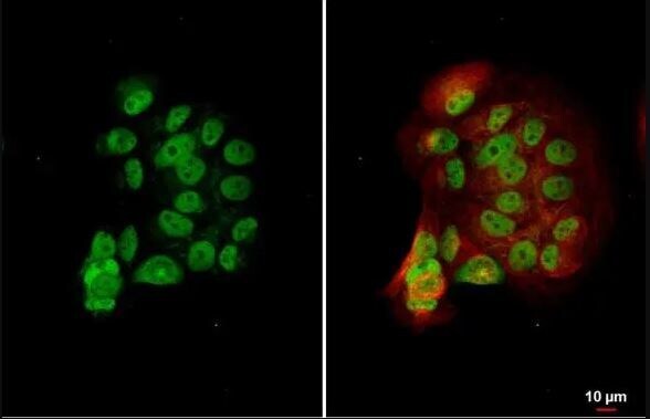
- Immunocytochemistry analysis of ESRRA in MCF-7 cells. Samples were incubated in ESRRA polyclonal antibody (Product # PA5-28749) using a dilution of 1:500. Sample: MCF-7 cells were fixed in 4% paraformaldehyde at RT for 15 min. Green: ERR alpha stained by ERR alpha antibody [N1], N-term. Red: alpha Tubulin, a cytoskeleton marker, stained by alpha Tubulin antibody [GT114] diluted at 1:1,000.

- Submitted by
- Invitrogen Antibodies (provider)
- Main image
- Experimental details
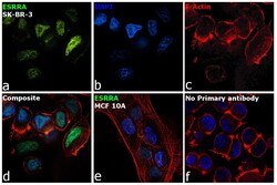
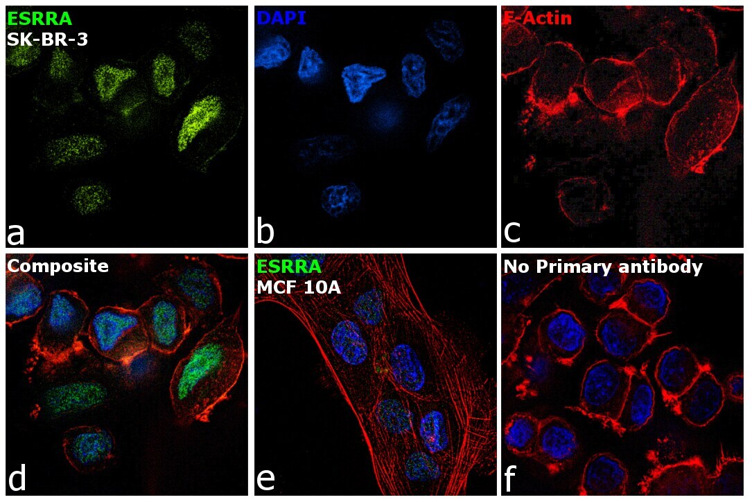
- Immunofluorescence analysis of ESRRA was performed using 70% confluent log phase SK-BR-3 and MCF 10A cells. The cells were fixed with 4% paraformaldehyde for 10 minutes, permeabilized with 0.1% Triton™ X-100 for 15 minutes, and blocked with 2% BSA for 1 hour at room temperature. The cells were labeled with ESRRA Polyclonal Antibody (Product # PA5-28749) at 1:100 dilution in 0.1% BSA, incubated at 4 degree celsius overnight and then with Goat anti-Rabbit IgG (H+L), Superclonal™ Recombinant Secondary Antibody, Alexa Fluor 488 conjugate (Product # A27034) at a dilution of 1:2000 for 45 minutes at room temperature (Panel a: green). Nuclei (Panel b: blue) were stained with SlowFade® Gold Antifade Mountant with DAPI (Product # S36938). F-actin (Panel c: red) was stained with Rhodamine Phalloidin (Product # R415, 1:300). Panel d represents the merged image showing predominantly nuclear localization. Panel e represents MCF 10A cells having low expression of ESRRA. Panel f represents control cells with no primary antibody to assess background. The images were captured at 60X magnification.
- Submitted by
- Invitrogen Antibodies (provider)
- Main image

- Experimental details
- Immunocytochemistry analysis of ESRRA in MCF-7 cells. Samples were incubated in ESRRA polyclonal antibody (Product # PA5-28749) using a dilution of 1:500. Sample: MCF-7 cells were fixed in 4% paraformaldehyde at RT for 15 min. Green: ERR alpha stained by ERR alpha antibody [N1], N-term. Red: alpha Tubulin, a cytoskeleton marker, stained by alpha Tubulin antibody [GT114] diluted at 1:1,000.
- Submitted by
- Invitrogen Antibodies (provider)
- Main image

- Experimental details
- Immunofluorescence analysis of ESRRA was performed using 70% confluent log phase SK-BR-3 and MCF 10A cells. The cells were fixed with 4% paraformaldehyde for 10 minutes, permeabilized with 0.1% Triton™ X-100 for 15 minutes, and blocked with 2% BSA for 1 hour at room temperature. The cells were labeled with ESRRA Polyclonal Antibody (Product # PA5-28749) at 1:100 dilution in 0.1% BSA, incubated at 4 degree celsius overnight and then with Goat anti-Rabbit IgG (Heavy Chain), Superclonal™ Recombinant Secondary Antibody, Alexa Fluor 488 conjugate (Product # A27034) at a dilution of 1:2000 for 45 minutes at room temperature (Panel a: green). Nuclei (Panel b: blue) were stained with SlowFade® Gold Antifade Mountant with DAPI (Product # S36938). F-actin (Panel c: red) was stained with Rhodamine Phalloidin (Product # R415, 1:300). Panel d represents the merged image showing predominantly nuclear localization. Panel e represents MCF 10A cells having low expression of ESRRA. Panel f represents control cells with no primary antibody to assess background. The images were captured at 60X magnification.

Supportive validation
- Submitted by
- Invitrogen Antibodies (provider)
- Main image
- Experimental details
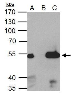
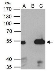
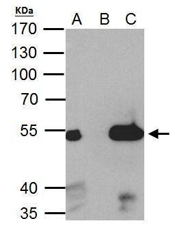
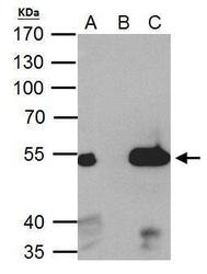
- ERR alpha antibody immunoprecipitates ERR alpha protein in IP experiments. IP Sample: 293T whole cell lysate/extract A. 40 µg 293T whole cell lysate/extract B. Control with 2 µg of preimmune rabbit IgG C. Immunoprecipitation of ERR alpha protein by 2 µg of ERR alpha antibody (Product # PA5-28749) 7.5% SDS-PAGE The immunoprecipitated ERR alpha protein was detected by ERR alpha antibody (Product # PA5-28749) diluted at 1:1,000.
Supportive validation
- Submitted by
- Invitrogen Antibodies (provider)
- Main image
- Experimental details
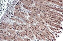
- Immunohistochemistry analysis of ESRRA in paraffin-embedded mouse stomach. Samples were incubated in ESRRA polyclonal antibody (Product # PA5-28749) using a dilution of 1:500. Antigen Retrieval: Citrate buffer, pH 6.0, 15 min.
- Submitted by
- Invitrogen Antibodies (provider)
- Main image
- Experimental details
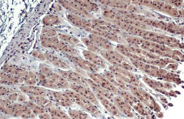
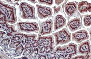
- Immunohistochemistry analysis of ESRRA in paraffin-embedded rat colon. Samples were incubated in ESRRA polyclonal antibody (Product # PA5-28749) using a dilution of 1:500. Antigen Retrieval: Citrate buffer, pH 6.0, 15 min.

- Submitted by
- Invitrogen Antibodies (provider)
- Main image

- Experimental details
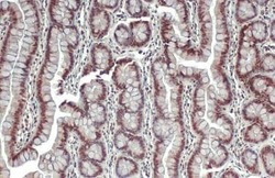
- Immunohistochemistry analysis of ESRRA in paraffin-embedded mouse intestine. Samples were incubated in ESRRA polyclonal antibody (Product # PA5-28749) using a dilution of 1:500. Antigen Retrieval: Citrate buffer, pH 6.0, 15 min.
Supportive validation
- Submitted by
- Invitrogen Antibodies (provider)
- Main image

- Experimental details
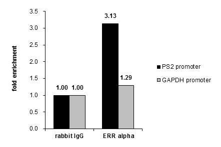
- Cross-linked ChIP was performed with MCF-7 chromatin extract and 5 µg of either control rabbit IgG or ESRRA Polyclonal Antibody (Product # PA5-28749). The precipitated DNA was detected by PCR with primer set targeting to PS2 promoter or GAPDH promoter.

- Submitted by
- Invitrogen Antibodies (provider)
- Main image

- Experimental details
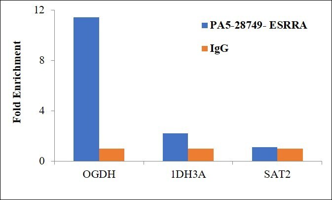
- Chromatin Immunoprecipitation (ChIP) assay of endogenous ESRRA protein using Anti-ESRRA Antibody: ChIP was performed using Anti-ESRRA Rabbit Polyclonal Antibody (Product # PA5-28749) 5 µg, on sheared chromatin from etoposide treated MCF-7 cells using the MAGnify ChIP System kit (Product # 49-2024). Normal Rabbit IgG was used as a negative IP control. The purified DNA was analyzed by qPCR using primers binding to promoter of OGDH, IDH3A promoter and SAT2 satellite repeats. Data is presented as fold enrichment of the antibody signal versus the negative control IgG using the comparative CT method.

- Submitted by
- Invitrogen Antibodies (provider)
- Main image

- Experimental details
- Cross-linked ChIP was performed with MCF-7 chromatin extract and 5 µg of either control rabbit IgG or ESRRA Polyclonal Antibody (Product # PA5-28749). The precipitated DNA was detected by PCR with primer set targeting to PS2 promoter or GAPDH promoter.
- Submitted by
- Invitrogen Antibodies (provider)
- Main image

- Experimental details
- Chromatin Immunoprecipitation (ChIP) assay of endogenous ESRRA protein using Anti-ESRRA Antibody: ChIP was performed using Anti-ESRRA Rabbit Polyclonal Antibody (Product # PA5-28749) 5 µg, on sheared chromatin from etoposide treated MCF-7 cells using the MAGnify ChIP System kit (Product # 49-2024). Normal Rabbit IgG was used as a negative IP control. The purified DNA was analyzed by qPCR using primers binding to promoter of OGDH, IDH3A promoter and SAT2 satellite repeats. Data is presented as fold enrichment of the antibody signal versus the negative control IgG using the comparative CT method.
Supportive validation
- Submitted by
- Invitrogen Antibodies (provider)
- Main image
- Experimental details
- ERR alpha antibody immunoprecipitates ERR alpha protein in IP experiments. IP Sample: 293T whole cell lysate/extract A. 40 µg 293T whole cell lysate/extract B. Control with 2 µg of preimmune rabbit IgG C. Immunoprecipitation of ERR alpha protein by 2 µg of ERR alpha antibody (Product # PA5-28749) 7.5% SDS-PAGE The immunoprecipitated ERR alpha protein was detected by ERR alpha antibody (Product # PA5-28749) diluted at 1:1,000.