Explore
 Validate
 Learn
LearnPA5-34509
antibody from Invitrogen Antibodies
Targeting: CCL4
Act-2, AT744.1, LAG1, MIP-1-beta, SCYA4
 Western blot
Antibody data
- Antibody Data
- Antigen structure
- References [1]
- Comments [0]
- Validations
- Western blot [1]
- Immunohistochemistry [2]
- Other assay [1]
Submit
Validation data
Reference
Comment
Report error
- Product number
- PA5-34509 - Provider product page

- Provider
- Invitrogen Antibodies
- Product name
- CCL4 Polyclonal Antibody
- Antibody type
- Polyclonal
- Antigen
- Synthetic peptide
- Description
- A suggested positive control is rat brain tissue lysate. PA5-34509 can be used with blocking peptide PEP-1552.
- Reactivity
- Human, Mouse, Rat
- Host
- Rabbit
- Isotype
- IgG
- Vial size
- 100 μg
- Concentration
- 1 mg/mL
- Storage
- Maintain refrigerated at 2-8°C for up to 3 months. For long term storage store at -20°C
Submitted references Vinpocetine alleviates lung inflammation via macrophage inflammatory protein-1β inhibition in an ovalbumin-induced allergic asthma model.
Choi WS, Kang HS, Kim HJ, Lee WT, Sohn UD, Lee JY
PloS one 2021;16(4):e0251012
PloS one 2021;16(4):e0251012
No comments: Submit comment
Supportive validation
- Submitted by
- Invitrogen Antibodies (provider)
- Main image
- Experimental details
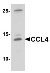
- Western Blot analysis of CCL4 in rat brain tissue lysate with CCL4 Polyclonal Antibody (Product # PA5-34509) at 1 µg/mL.

Supportive validation
- Submitted by
- Invitrogen Antibodies (provider)
- Main image
- Experimental details
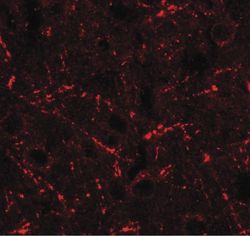
- Immunofluorescence of CCL4 in rat brain tissue with CCL4 Polyclonal Antibody (Product # PA5-34509) at 20 µg/mL.

- Submitted by
- Invitrogen Antibodies (provider)
- Main image
- Experimental details
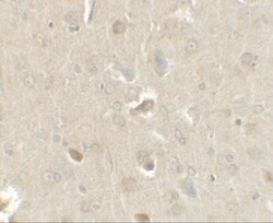
- Immunohistochemistry of CCL4 in rat brain tissue with CCL4 Polyclonal Antibody (Product # PA5-34509) at 2.5 µg/mL.

Supportive validation
- Submitted by
- Invitrogen Antibodies (provider)
- Main image

- Experimental details
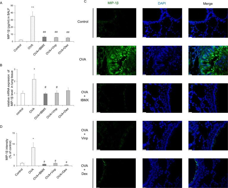
- Fig 4 Effects of IBMX and Vinp on MIP-1beta expression and release in eosinophilic lung inflammation. ( A ) MIP-1beta in BALF was measured by ELISA. ( B ) The mRNA expression level of MIP-1beta in lung tissues was measured by RT-qPCR. ( C ) MIP-1beta expression in lung tissue was detected by immunofluorescence staining (magnification, 63x; scale bar, 20 mum). ( D ) In immunofluorescence-stained tissue, the green fluorescence intensity of stained images was measured using ImageJ. Data are expressed as mean +- SEM. Statistical analysis was performed using one-way ANOVA (Data were considered significant at ** P