Explore
 Validate
 Learn
Learn Western blot
 Immunocytochemistry
Antibody data
- Antibody Data
- Antigen structure
- References [2]
- Comments [0]
- Validations
- Immunocytochemistry [2]
- Flow cytometry [1]
- Chromatin Immunoprecipitation [2]
- Other assay [1]
Submit
Validation data
Reference
Comment
Report error
- Product number
- 720259 - Provider product page

- Provider
- Invitrogen Antibodies
- Product name
- TCF2 Polyclonal Antibody
- Antibody type
- Polyclonal
- Antigen
- Synthetic peptide
- Description
- These Polyclonal antibodies are of rabbit origin developed by immunizing animals with proteins or peptides. The polyclonal antibody is purified by affinity purification from the rabbit sera generated after immunizing the rabbits with a specific type of protein or peptide. The purified antibody is tested for its functionality in various relevant research applications. The antibody is developed for Research Use Only and is non-hazardous or non-infectious in nature. This antibody is predicted to react with Monkey, Horse and Cat.
- Reactivity
- Human, Mouse, Rat
- Host
- Rabbit
- Isotype
- IgG
- Vial size
- 100 μg
- Concentration
- 0.5 mg/mL
- Storage
- Store at 4°C short term. For long term storage, store at -20°C, avoiding freeze/thaw cycles.
Submitted references An HNF1α truncation associated with maturity-onset diabetes of the young impairs pancreatic progenitor differentiation by antagonizing HNF1β function.
Enhancer and super-enhancer dynamics in repair after ischemic acute kidney injury.
Cujba AM, Alvarez-Fallas ME, Pedraza-Arevalo S, Laddach A, Shepherd MH, Hattersley AT, Watt FM, Sancho R
Cell reports 2022 Mar 1;38(9):110425
Cell reports 2022 Mar 1;38(9):110425
Enhancer and super-enhancer dynamics in repair after ischemic acute kidney injury.
Wilflingseder J, Willi M, Lee HK, Olauson H, Jankowski J, Ichimura T, Erben R, Valerius MT, Hennighausen L, Bonventre JV
Nature communications 2020 Jul 7;11(1):3383
Nature communications 2020 Jul 7;11(1):3383
No comments: Submit comment
Supportive validation
- Submitted by
- Invitrogen Antibodies (provider)
- Main image

- Experimental details
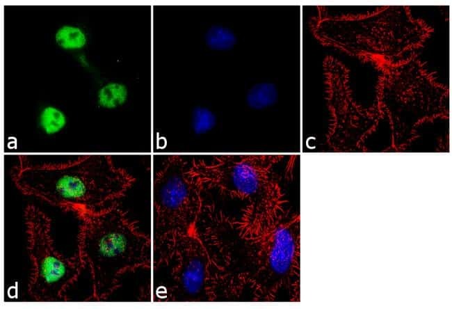
- For immunofluorescence analysis, Hep G2 cells were fixed and permeabilized for detection of endogenous HNF1b using HNF1b Rabbit Polyclonal Antibody (Product# 720259, 2 µg/mL) and labeled with Goat anti-Rabbit IgG (H+L) Superclonal Secondary Antibody, Alexa Fluor® 488 conjugate (Product # A27034, 1:2000). Panel a) shows representative cells that were stained for detection and localization of HNF1b protein (green), Panel b) is stained for nuclei (blue) using SlowFade® Gold Antifade Mountant with DAPI (Product # S36938). Panel c) represents cytoskeletal F-actin staining using Rhodamine Phalloidin (Product # R415, 1:300). Panel d) is a composite image of Panels a, b and c clearly demonstrating nuclear localization of HNF1b. Panel e)represents control cells with no primary antibody to assess background. The images were captured at 60X magnification.
- Submitted by
- Invitrogen Antibodies (provider)
- Main image
- Experimental details
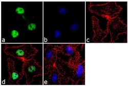
- For immunofluorescence analysis, Hep G2 cells were fixed and permeabilized for detection of endogenous HNF1b using HNF1b Rabbit Polyclonal Antibody (Product# 720259, 2 µg/mL) and labeled with Goat anti-Rabbit IgG (Heavy Chain) Superclonal Secondary Antibody, Alexa Fluor® 488 conjugate (Product # A27034, 1:2000). Panel a) shows representative cells that were stained for detection and localization of HNF1b protein (green), Panel b) is stained for nuclei (blue) using SlowFade® Gold Antifade Mountant with DAPI (Product # S36938). Panel c) represents cytoskeletal F-actin staining using Rhodamine Phalloidin (Product # R415, 1:300). Panel d) is a composite image of Panels a, b and c clearly demonstrating nuclear localization of HNF1b. Panel e)represents control cells with no primary antibody to assess background. The images were captured at 60X magnification.

Supportive validation
- Submitted by
- Invitrogen Antibodies (provider)
- Main image
- Experimental details
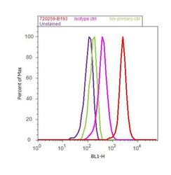
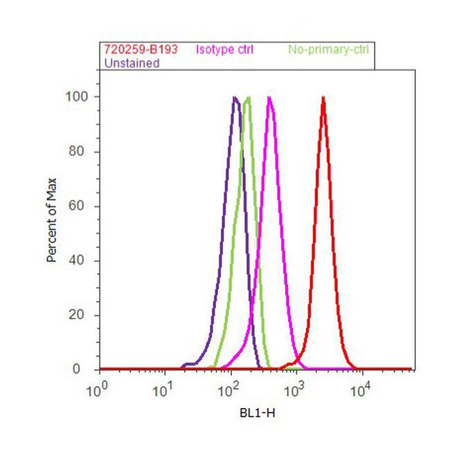
- Flow Cytometry analysis of endogenous HNF1b was performed on K-562 cells labeled with Anti-HNF1b Rabbit Polyclonal Antibody (Product# 720259, 5 ug/ 1M cells) or with Rabbit isotype control and detected with Goat anti-Rabbit IgG (H+L) Superclonal™ Secondary Antibody, (Alexa Fluor® 488 conjugate, Product# A27034, 0.4 ug/ml, 1:2500) as represented by the red and pink histograms respectively. The purple histogram represents unstained control cells and the green histogram represents no-primary-Antibody control. A representative of 10,000 cells were acquired and analyzed for each sample using an Attune® Acoustic Focusing Cytometer (4468770).
Supportive validation
- Submitted by
- Invitrogen Antibodies (provider)
- Main image
- Experimental details
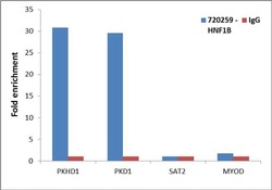
- Enrichment of endogenous HNF1B protein at specific gene loci using Anti-HNF1B Rabbit Polyclonal Antibody: Chromatin Immunoprecipitation (ChIP) was performed using Anti-HNF1B Rabbit Polyclonal Antibody (Product # 720259, 3 µg) on sheared chromatin from 2 million HEPG2 cells using the MAGnify ChIP system kit (Product # 49-2024). Normal Rabbit IgG (1 µg) was used as a negative IP control. The purified DNA was analyzed by 7500 Fast qPCR system (Product # 4351106) with optimized PCR primer pairs for the promoters of the active PKHD1, PKD1 region used as positive control target genes, and the region of the inactive MYOD, SAT2 satellite repeat, used as negative control target gene. Data is presented as fold enrichment of the antibody signal versus the negative control IgG using the comparative CT method.

- Submitted by
- Invitrogen Antibodies (provider)
- Main image
- Experimental details
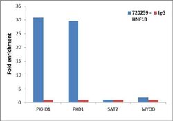
- Enrichment of endogenous HNF1B protein at specific gene loci using Anti-HNF1B Rabbit Polyclonal Antibody: Chromatin Immunoprecipitation (ChIP) was performed using Anti-HNF1B Rabbit Polyclonal Antibody (Product # 720259, 3 µg) on sheared chromatin from 2 million HEPG2 cells using the MAGnify ChIP system kit (Product # 49-2024). Normal Rabbit IgG (1 µg) was used as a negative IP control. The purified DNA was analyzed by 7500 Fast qPCR system (Product # 4351106) with optimized PCR primer pairs for the promoters of the active PKHD1, PKD1 region used as positive control target genes, and the region of the inactive MYOD, SAT2 satellite repeat, used as negative control target gene. Data is presented as fold enrichment of the antibody signal versus the negative control IgG using the comparative CT method.

Supportive validation
- Submitted by
- Invitrogen Antibodies (provider)
- Main image

- Experimental details
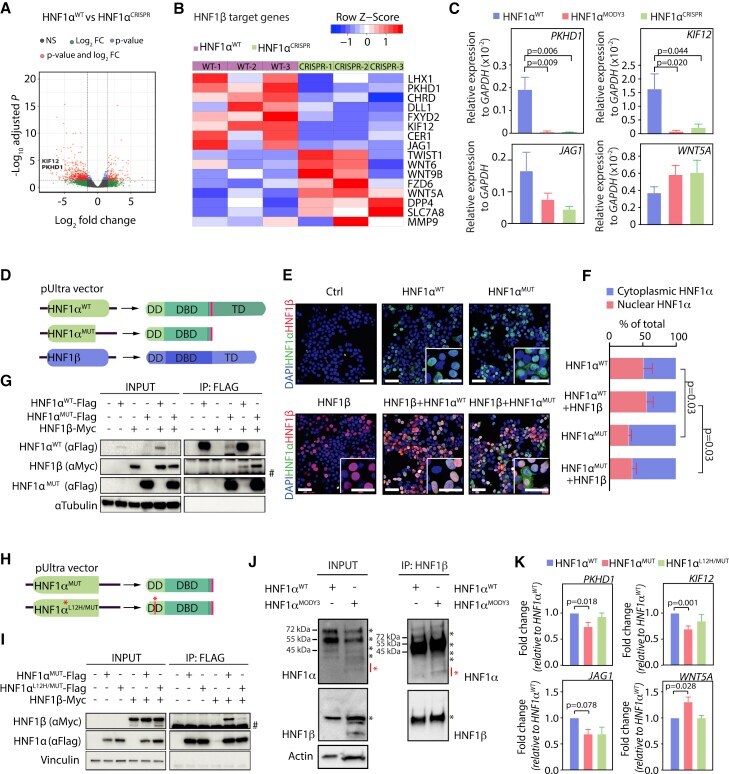
- Figure 5 HNF1alpha p291fsinsC truncated protein interacts with HNF1beta and impairs HNF1b-dependent transcription (A) Volcano plot of significantly differentially expressed genes in HNF1alpha WT versus HNF1alpha CRISPR PPs. PKHD1 and KIF12 are indicated. (B) Heatmap of differentially expressed HNF1beta targets in HNF1alpha WT /HNF1alpha CRISPR PPs. (C) qRT-PCR analysis of HNF1beta target genes PKHD1 , KIF12 , TWIST1 , and WNT5A . n = 3 independent differentiations. (D) Schematic depicting the constructs used for overexpression of HNF1alpha WT /HNF1alpha MUT and HNF1beta proteins. (E) IF analysis on HNF1alpha/HNF1beta transfected HEK293T cells. (F) Nuclear/cytoplasmic HNF1alpha fluorescence intensity quantification in HEK293 transfected with HNF1alpha WT /HNF1alpha MUT +- HNF1beta. (G) FLAG immunoprecipitation in HNF1alpha/HNF1beta transfected HEK293T cells. Vinculin, HNF1beta, and HNF1alpha were detected by immunoblot in the input and immunoprecipitation (IP). n = 3 independent experiments. (H) Schematic depicting the constructs used for HNF1alpha MUT /HNF1alpha L12H/MUT overexpression. Red asterisk depicts the point mutation L12H known to affect HNF1alpha dimerization domain. (I) FLAG immunoprecipitation in HNF1alpha/HNF1beta transfected HEK293T cells. Vinculin, HNF1beta, and HNF1alpha were detected by immunoblot in the input and IP. n = 3 independent experiments. (J) HNF1beta immunoprecipitation in HNF1alpha WT /HNF1alpha MODY3 PP organoids. HNF1beta and HNF1alpha were dete