Explore
 Validate
 Learn
Learn Western blot
 Immunocytochemistry
Antibody data
- Antibody Data
- Antigen structure
- References [1]
- Comments [0]
- Validations
- Immunocytochemistry [2]
- Immunohistochemistry [6]
Submit
Validation data
Reference
Comment
Report error
- Product number
- MA5-24605 - Provider product page

- Provider
- Invitrogen Antibodies
- Product name
- TCF2 Monoclonal Antibody (CL0374)
- Antibody type
- Monoclonal
- Antigen
- Recombinant full-length protein
- Description
- Immunogen sequence: KEVLVQALEE LLPSPNFGVK LETLPLSPGS GAEPDTKPVF HTLTNGHAKG RLSGDEGSED GDDYDTPPIL KELQALNTEE AAEQRAEVDR MLSEDPWRAA KMIKGYMQQH Highest antigen sequence identity to the following orthologs: Mouse - 97%, Rat - 97%. Binds to an epitope located within the peptide sequence TPPILKELQA as determined by overlapping synthetic peptides.
- Reactivity
- Human
- Host
- Mouse
- Isotype
- IgG
- Antibody clone number
- CL0374
- Vial size
- 100 μL
- Concentration
- 1 mg/mL
- Storage
- Store at 4°C short term. For long term storage, store at -20°C, avoiding freeze/thaw cycles.
Submitted references Utility of HNF-1B and a panel of lineage-specific biomarkers to optimize the diagnosis of pancreatic ductal adenocarcinoma.
Bai S, Lindberg J, Whalen G, Bathini V, Zou J, Yang MX
American journal of cancer research 2021;11(3):858-865
American journal of cancer research 2021;11(3):858-865
No comments: Submit comment
Supportive validation
- Submitted by
- Invitrogen Antibodies (provider)
- Main image

- Experimental details
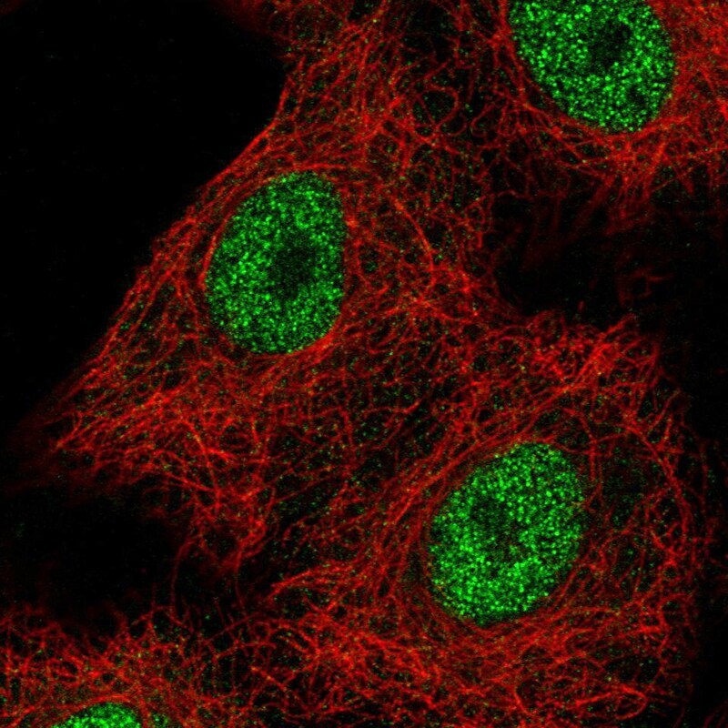
- Immunofluorescence staining of TCF2 in A549 cell line using TCF2 Monoclonal Antibody (Product # MA5-24605), showing spotty nuclear (without nucleoli) staining in green. Microtubule probes are visualized in red (where available).

- Submitted by
- Invitrogen Antibodies (provider)
- Main image

- Experimental details
- Immunofluorescent analysis of TCF2 in A549 cell line using TCF2 Monoclonal Antibody (CL0374) (Product # MA5-24605). Staining shows spotty nuclear (without nucleoli) staining in green. Microtubule probes are visualized in red (where available).
Supportive validation
- Submitted by
- Invitrogen Antibodies (provider)
- Main image

- Experimental details
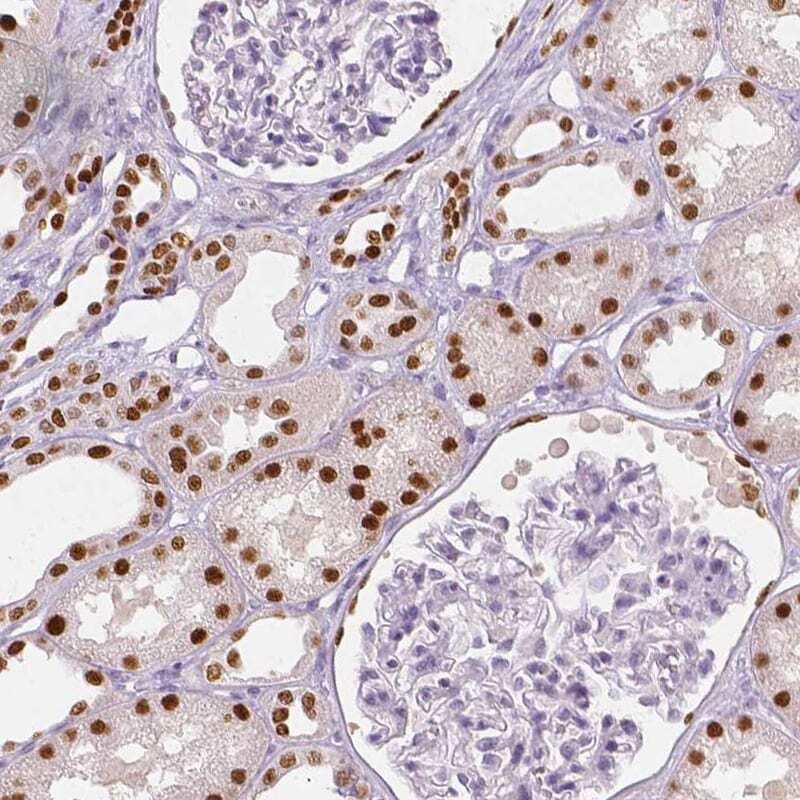
- Immunohistochemical staining of TCF2 in human kidney using TCF2 Monoclonal Antibody (CL0374) (Product # MA5-24605) shows strong nuclear positivity in cells in tubules.
- Submitted by
- Invitrogen Antibodies (provider)
- Main image
- Experimental details
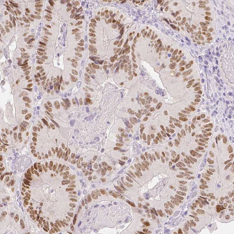
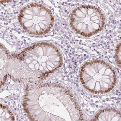
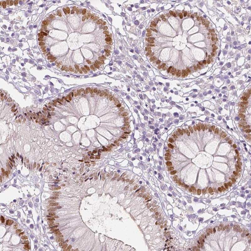
- Immunohistochemical staining of TCF2 in human colon using TCF2 Monoclonal Antibody (CL0374) (Product # MA5-24605) shows moderate to strong nuclear positivity in glandular cells.
- Submitted by
- Invitrogen Antibodies (provider)
- Main image
- Experimental details
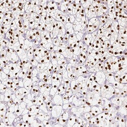
- Immunohistochemical staining of TCF2 in human renal cancer using TCF2 Monoclonal Antibody (CL0374) (Product # MA5-24605) shows strong nuclear positivity in tumor cells.
- Submitted by
- Invitrogen Antibodies (provider)
- Main image
- Experimental details
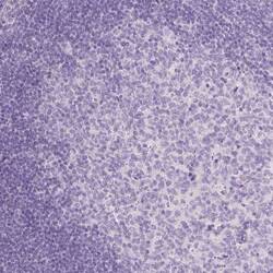
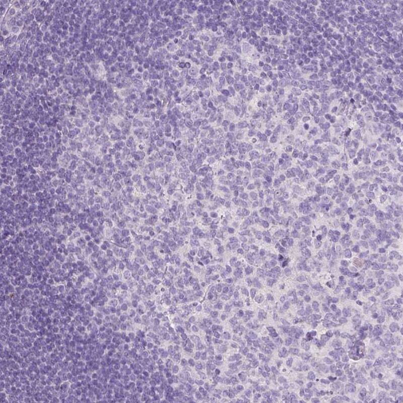
- Immunohistochemical staining of TCF2 in human tonsil using TCF2 Monoclonal Antibody (CL0374) (Product # MA5-24605) shows no positivity in lymphoid cells as expected.
- Submitted by
- Invitrogen Antibodies (provider)
- Main image
- Experimental details
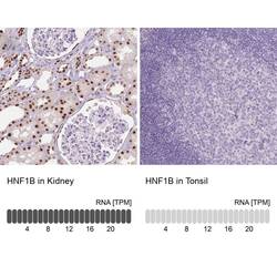
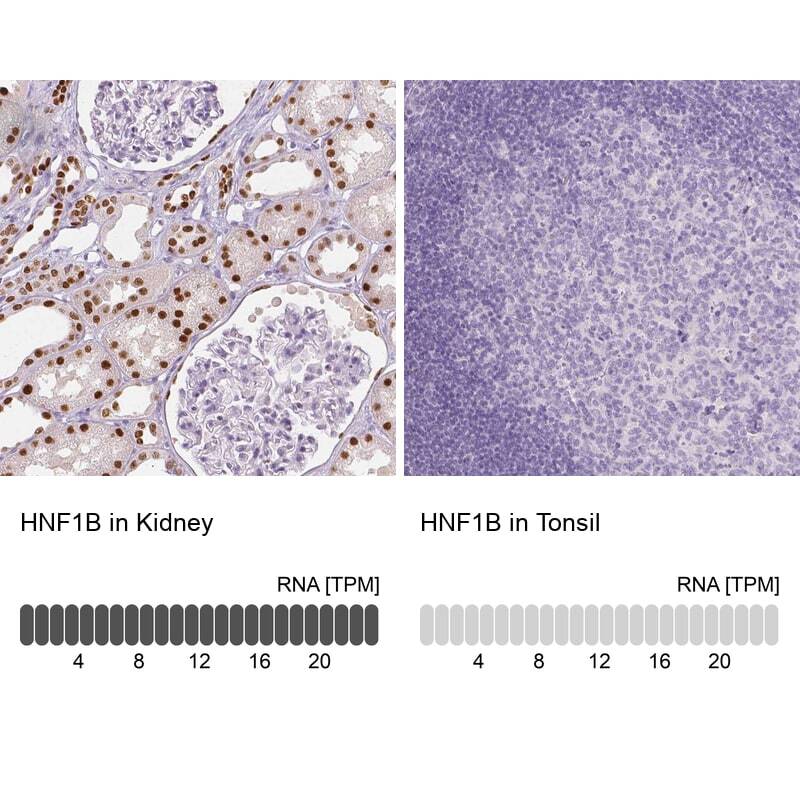
- Immunohistochemical staining of TCF2 in human kidney and tonsil tissues using TCF2 Monoclonal Antibody (CL0374) (Product # MA5-24605). Corresponding HNF1B RNA-seq data are presented for the same tissues.
- Submitted by
- Invitrogen Antibodies (provider)
- Main image

- Experimental details
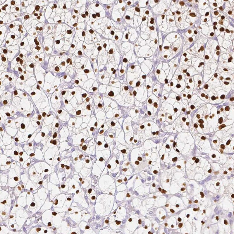
- Immunohistochemical staining of TCF2 in human colorectal cancer using TCF2 Monoclonal Antibody (CL0374) (Product # MA5-24605) shows moderate nuclear positivity in tumor cells.