 Explore
 Validate
 Learn
Learn Western blot
 Immunocytochemistry
Antibody data
- Antibody Data
- Antigen structure
- References [5]
- Comments [0]
- Validations
- Western blot [5]
- Immunohistochemistry [6]
Submit
Validation data
Reference
Comment
Report error
- Product number
- NBP1-89680 - Provider product page

- Provider
- Novus Biologicals
- Proper citation
- Novus Cat#NBP1-89680, RRID:AB_11035007
- Product name
- Rabbit Polyclonal TCF-2/HNF-1 beta Antibody
- Antibody type
- Polyclonal
- Description
- Immunogen affinity purified. Specificity of human TCF-2/HNF-1 beta antibody verified on a Protein Array containing target protein plus 383 other non-specific proteins.
- Reactivity
- Human, Mouse
- Host
- Rabbit
- Isotype
- IgG
- Vial size
- 0.1 ml
- Storage
- Store at 4C short term. Aliquot and store at -20C long term. Avoid freeze-thaw cycles.
Submitted references Obesity-induced reduced expression of the lncRNA ROIT impairs insulin transcription by downregulation of Nkx6.1 methylation.
Long-term primary culture of a clear cell ovarian carcinoma reveals an epithelial-mesenchymal cooperative interaction.
Hepatocyte nuclear factor-1β is not a specific marker of clear cell carcinoma in serous effusions.
Role of PINCH and its partner tumor suppressor Rsu-1 in regulating liver size and tumorigenesis.
Genomic profiling of papillary renal cell tumours identifies small regions of DNA alterations: a possible role of HNF1B in tumour development.
Zhang FF, Liu YH, Wang DW, Liu TS, Yang Y, Guo JM, Pan Y, Zhang YF, Du H, Li L, Jin L
Diabetologia 2020 Apr;63(4):811-824
Diabetologia 2020 Apr;63(4):811-824
Long-term primary culture of a clear cell ovarian carcinoma reveals an epithelial-mesenchymal cooperative interaction.
Goyeneche AA, Koch M, Bell MC, Telleria CM
Cancer cell international 2015;15:88
Cancer cell international 2015;15:88
Hepatocyte nuclear factor-1β is not a specific marker of clear cell carcinoma in serous effusions.
Davidson B
Cancer cytopathology 2014 Feb;122(2):153-8
Cancer cytopathology 2014 Feb;122(2):153-8
Role of PINCH and its partner tumor suppressor Rsu-1 in regulating liver size and tumorigenesis.
Donthamsetty S, Bhave VS, Mars WM, Bowen WC, Orr A, Haynes MM, Wu C, Michalopoulos GK
PloS one 2013;8(9):e74625
PloS one 2013;8(9):e74625
Genomic profiling of papillary renal cell tumours identifies small regions of DNA alterations: a possible role of HNF1B in tumour development.
Szponar A, Yusenko MV, Kuiper R, van Kessel AG, Kovacs G
Histopathology 2011 May;58(6):934-43
Histopathology 2011 May;58(6):934-43
No comments: Submit comment
Supportive validation
- Submitted by
- Novus Biologicals (provider)
- Main image

- Experimental details
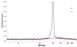
- Simple Western: TCF-2/HNF-1 beta Antibody [NBP1-89680] - Simple Western lane view shows a specific band for HNF1B in 0.2 mg/ml of A549 lysate. This experiment was performed under reducing conditions using the 12-230 kDa separation system.

- Submitted by
- Novus Biologicals (provider)
- Main image
- Experimental details
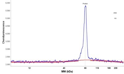
- Simple Western: TCF-2/HNF-1 beta Antibody [NBP1-89680] - Electropherogram image(s) of corresponding Simple Western lane view. TCF-2/HNF-1 beta antibody was used at 1:30 dilution on A549 lysate(s).
- Submitted by
- Novus Biologicals (provider)
- Main image

- Experimental details
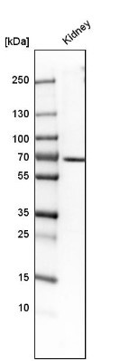
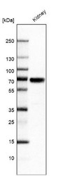
- Western Blot: TCF-2/HNF-1 beta Antibody [NBP1-89680] - Analysis in human kidney tissue.
- Submitted by
- Novus Biologicals (provider)
- Main image

- Experimental details
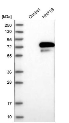
- Western Blot: TCF-2/HNF-1 beta Antibody [NBP1-89680] - Analysis in control (vector only transfected HEK293T lysate) and HNF1B over-expression lysate (Co-expressed with a C-terminal myc-DDK tag (3.1 kDa) in mammalian HEK293T cells).
- Submitted by
- Novus Biologicals (provider)
- Main image
- Experimental details
- Western Blot: TCF-2/HNF-1 beta Antibody [NBP1-89680] - Analysis in human kidney tissue.

Supportive validation
- Submitted by
- Novus Biologicals (provider)
- Main image

- Experimental details
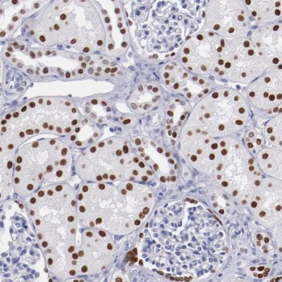
- Immunohistochemistry-Paraffin: TCF-2/HNF-1 beta Antibody [NBP1-89680] - Staining of human kidney shows moderate to strong nuclear positivity in renal tubules.
- Submitted by
- Novus Biologicals (provider)
- Main image
- Experimental details
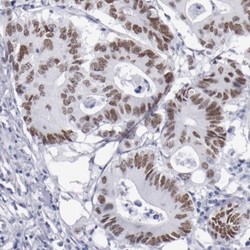
- Immunohistochemistry-Paraffin: TCF-2/HNF-1 beta Antibody [NBP1-89680] - Staining of human colorectal cancer shows moderate nuclear positivity in tumor cells.

- Submitted by
- Novus Biologicals (provider)
- Main image
- Experimental details
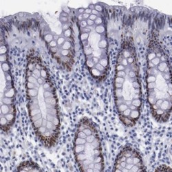
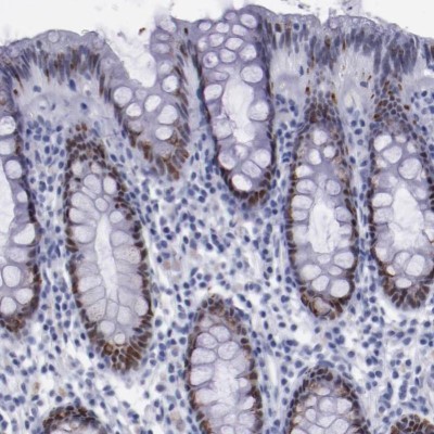
- Immunohistochemistry-Paraffin: TCF-2/HNF-1 beta Antibody [NBP1-89680] - Staining of human rectum shows strong nuclear positivity in glandular cells.
- Submitted by
- Novus Biologicals (provider)
- Main image
- Experimental details
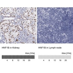
- Immunohistochemistry-Paraffin: TCF-2/HNF-1 beta Antibody [NBP1-89680] - Analysis in human kidney and lymph node tissues. Corresponding HNF1B RNA-seq data are presented for the same tissues.

- Submitted by
- Novus Biologicals (provider)
- Main image
- Experimental details
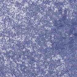
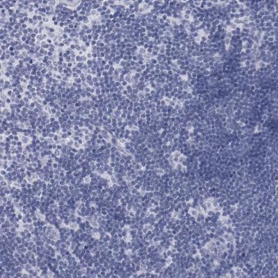
- Immunohistochemistry-Paraffin: TCF-2/HNF-1 beta Antibody [NBP1-89680] - Staining of human lymph node shows no positivity in lymphoid cells as expected.
- Submitted by
- Novus Biologicals (provider)
- Main image
- Experimental details
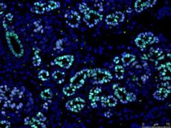
- Immunohistochemistry-Paraffin: TCF-2/HNF-1 beta Antibody [NBP1-89680] - Nuclear expression of HNF1B (green) in a subset of PDX1 expressing cells (blue) in human pancreas. Image submitted by a verified customer review.
