Explore
 Validate
 Learn
Learn Western blot
 Immunocytochemistry
Antibody data
- Antibody Data
- Antigen structure
- References [22]
- Comments [0]
- Validations
- Immunocytochemistry [1]
- Immunohistochemistry [1]
Submit
Validation data
Reference
Comment
Report error
- Product number
- HPA002083 - Provider product page

- Provider
- Atlas Antibodies
- Proper citation
- Atlas Antibodies Cat#HPA002083, RRID:AB_1080232
- Product name
- Anti-HNF1B
- Antibody type
- Polyclonal
- Description
- Polyclonal Antibody against Human HNF1B, Gene description: HNF1 homeobox B, Alternative Gene Names: HNF1beta, LFB3, MODY5, TCF2, VHNF1, Validated applications: WB, IHC, ICC, Uniprot ID: P35680, Storage: Store at +4°C for short term storage. Long time storage is recommended at -20°C.
- Reactivity
- Human
- Host
- Rabbit
- Conjugate
- Unconjugated
- Isotype
- IgG
- Vial size
- 100 µl
- Concentration
- 0.3 mg/ml
- Storage
- Store at +4°C for short term storage. Long time storage is recommended at -20°C.
- Handling
- The antibody solution should be gently mixed before use.
Submitted references Cell State of Origin Impacts Development of Distinct Endometriosis-Related Ovarian Carcinoma Histotypes
Identification of a ΔNp63-Dependent Basal-Like A Subtype-Specific Transcribed Enhancer Program (B-STEP) in Aggressive Pancreatic Ductal Adenocarcinoma
Establishment and characterization of a novel ovarian high-grade serous carcinoma cell line—IPO43
HNF1B‐driven three‐dimensional chromatin structure for molecular classification in pancreatic cancers
The Expression of Transcription Factors in Fetal Lamb Kidney
Endometrial Tumor Classification by Histomorphology and Biomarkers in the Nurses’ Health Study
Pancreatic cancer prognosis is predicted by an ATAC-array technology for assessing chromatin accessibility
Napsin-A and AMACR are Superior to HNF-1β in Distinguishing Between Mesonephric Carcinomas and Clear Cell Carcinomas of the Gynecologic Tract
Analysis of expression, epigenetic, and genetic changes of HNF1B in 130 kidney tumours
Protocol for Large-Scale Production of Kidney Organoids from Human Pluripotent Stem Cells
FOXA 2 controls the cis ‐regulatory networks of pancreatic cancer cells in a differentiation grade‐specific manner
Characterization and drug sensitivity of a novel human ovarian clear cell carcinoma cell line genomically and phenotypically similar to the original tumor
Investigation of HNF-1B as a diagnostic biomarker for pancreatic ductal adenocarcinoma
Caudal migration and proliferation of renal progenitors regulates early nephron segment size in zebrafish
Molecular Analysis of Mixed Endometrial Carcinomas Shows Clonality in Most Cases
Dissection of transcriptional and cis ‐regulatory control of differentiation in human pancreatic cancer
Prognostic relevance of molecular subtypes and master regulators in pancreatic ductal adenocarcinoma
Hepatocyte Nuclear Factor-1β Induces Redifferentiation of Dedifferentiated Tubular Epithelial Cells
Long-term primary culture of a clear cell ovarian carcinoma reveals an epithelial–mesenchymal cooperative interaction
Role of PINCH and Its Partner Tumor Suppressor Rsu-1 in Regulating Liver Size and Tumorigenesis
Hepatocyte nuclear factor‐1β is not a specific marker of clear cell carcinoma in serous effusions
Genomic profiling of papillary renal cell tumours identifies small regions of DNA alterations: a possible role of HNF1B in tumour development
Beddows I, Fan H, Heinze K, Johnson B, Leonova A, Senz J, Djirackor S, Cho K, Pearce C, Huntsman D, Anglesio M, Shen H
Cancer Research 2024;84(1):26-38
Cancer Research 2024;84(1):26-38
Identification of a ΔNp63-Dependent Basal-Like A Subtype-Specific Transcribed Enhancer Program (B-STEP) in Aggressive Pancreatic Ductal Adenocarcinoma
Wang X, Kutschat A, Aggrey-Fynn J, Hamdan F, Graham R, Wixom A, Souto Y, Ladigan-Badura S, Yonkus J, Abdelrahman A, Alva-Ruiz R, Gaedcke J, Ströbel P, Kosinsky R, Wegwitz F, Hermann P, Truty M, Siveke J, Hahn S, Hessmann E, Johnsen S, Najafova Z
Molecular Cancer Research 2023;21(9):881-891
Molecular Cancer Research 2023;21(9):881-891
Establishment and characterization of a novel ovarian high-grade serous carcinoma cell line—IPO43
Silva F, Coelho F, Peixoto A, Pinto P, Martins C, Frombach A, Santo V, Brito C, Guimarães A, Félix A
Cancer Cell International 2022;22(1)
Cancer Cell International 2022;22(1)
HNF1B‐driven three‐dimensional chromatin structure for molecular classification in pancreatic cancers
Kato H, Tateishi K, Iwadate D, Yamamoto K, Fujiwara H, Nakatsuka T, Kudo Y, Hayakawa Y, Ijichi H, Otsuka M, Kishikawa T, Takahashi R, Miyabayashi K, Nakai Y, Hirata Y, Toyoda A, Morishita S, Fujishiro M
Cancer Science 2022;114(4):1672-1685
Cancer Science 2022;114(4):1672-1685
The Expression of Transcription Factors in Fetal Lamb Kidney
Nishiya Y, Kawaguchi K, Kudo K, Kawaguchi T, Obayashi J, Tanaka K, Ohyama K, Nagae H, Furuta S, Seki Y, Koike J, Pringle K, Kitagawa H
Journal of Developmental Biology 2021;9(2):22
Journal of Developmental Biology 2021;9(2):22
Endometrial Tumor Classification by Histomorphology and Biomarkers in the Nurses’ Health Study
Watkins J, Downing M, Crous-Bou M, Busch E, Chen M, De Vivo I, Mutter G, Antwi S
Journal of Cancer Epidemiology 2021;2021
Journal of Cancer Epidemiology 2021;2021
Pancreatic cancer prognosis is predicted by an ATAC-array technology for assessing chromatin accessibility
Dhara S, Chhangawala S, Chintalapudi H, Askan G, Aveson V, Massa A, Zhang L, Torres D, Makohon-Moore A, Lecomte N, Melchor J, Bermeo J, Cardenas A, Sinha S, Glassman D, Nicolle R, Moffitt R, Yu K, Leppanen S, Laderman S, Curry B, Gui J, Balachandran V, Iacobuzio-Donahue C, Chandwani R, Leslie C, Leach S
Nature Communications 2021;12(1)
Nature Communications 2021;12(1)
Napsin-A and AMACR are Superior to HNF-1β in Distinguishing Between Mesonephric Carcinomas and Clear Cell Carcinomas of the Gynecologic Tract
Pors J, Segura S, Cheng A, Ji J, Tessier-Cloutier B, Cochrane D, Fix D, Park K, Gilks B, Hoang L
Applied Immunohistochemistry & Molecular Morphology 2020;28(8):593-601
Applied Immunohistochemistry & Molecular Morphology 2020;28(8):593-601
Analysis of expression, epigenetic, and genetic changes of HNF1B in 130 kidney tumours
Bártů M, Hojný J, Hájková N, Michálková R, Krkavcová E, Hadravský L, Kleissnerová L, Bui Q, Stružinská I, Němejcová K, Čapoun O, Šlemendová M, Dundr P
Scientific Reports 2020;10(1)
Scientific Reports 2020;10(1)
Protocol for Large-Scale Production of Kidney Organoids from Human Pluripotent Stem Cells
Sander V, Przepiorski A, Crunk A, Hukriede N, Holm T, Davidson A
STAR Protocols 2020;1(3):100150
STAR Protocols 2020;1(3):100150
FOXA 2 controls the cis ‐regulatory networks of pancreatic cancer cells in a differentiation grade‐specific manner
Milan M, Balestrieri C, Alfarano G, Polletti S, Prosperini E, Spaggiari P, Zerbi A, Diaferia G, Natoli G
The EMBO Journal 2019;38(20)
The EMBO Journal 2019;38(20)
Characterization and drug sensitivity of a novel human ovarian clear cell carcinoma cell line genomically and phenotypically similar to the original tumor
Franklin M, Gentles L, Matheson E, Bown N, Cross P, Ralte A, Gilkes‐Immeson C, Bradbury A, Zanjirband M, Lunec J, Drew Y, O'Donnell R, Curtin N
Cancer Medicine 2018;7(9):4744-4754
Cancer Medicine 2018;7(9):4744-4754
Investigation of HNF-1B as a diagnostic biomarker for pancreatic ductal adenocarcinoma
Yang M, Coates R, Ambaye A, Gardner J, Zubarick R, Gao Y, Skelly J, Liu J, Mino-Kenudson M
Biomarker Research 2018;6(1)
Biomarker Research 2018;6(1)
Caudal migration and proliferation of renal progenitors regulates early nephron segment size in zebrafish
Naylor R, Dodd R, Davidson A
Scientific Reports 2016;6(1)
Scientific Reports 2016;6(1)
Molecular Analysis of Mixed Endometrial Carcinomas Shows Clonality in Most Cases
Köbel M, Meng B, Hoang L, Almadani N, Li X, Soslow R, Gilks C, Lee C
American Journal of Surgical Pathology 2016;40(2):166-180
American Journal of Surgical Pathology 2016;40(2):166-180
Dissection of transcriptional and cis ‐regulatory control of differentiation in human pancreatic cancer
Diaferia G, Balestrieri C, Prosperini E, Nicoli P, Spaggiari P, Zerbi A, Natoli G
The EMBO Journal 2016;35(6):595-617
The EMBO Journal 2016;35(6):595-617
Prognostic relevance of molecular subtypes and master regulators in pancreatic ductal adenocarcinoma
Janky R, Binda M, Allemeersch J, Van den broeck A, Govaere O, Swinnen J, Roskams T, Aerts S, Topal B
BMC Cancer 2016;16(1)
BMC Cancer 2016;16(1)
Hepatocyte Nuclear Factor-1β Induces Redifferentiation of Dedifferentiated Tubular Epithelial Cells
Dussaule J, Omata M, Doke Y, Yamada C, Kawashima K, Sho R, Enomoto K, Furuya M, Inomata N
PLOS ONE 2016;11(5):e0154912
PLOS ONE 2016;11(5):e0154912
Long-term primary culture of a clear cell ovarian carcinoma reveals an epithelial–mesenchymal cooperative interaction
Goyeneche A, Koch M, Bell M, Telleria C
Cancer Cell International 2015;15(1)
Cancer Cell International 2015;15(1)
Role of PINCH and Its Partner Tumor Suppressor Rsu-1 in Regulating Liver Size and Tumorigenesis
Singh S, Donthamsetty S, Bhave V, Mars W, Bowen W, Orr A, Haynes M, Wu C, Michalopoulos G
PLoS ONE 2013;8(9):e74625
PLoS ONE 2013;8(9):e74625
Hepatocyte nuclear factor‐1β is not a specific marker of clear cell carcinoma in serous effusions
Davidson B
Cancer Cytopathology 2013;122(2):153-158
Cancer Cytopathology 2013;122(2):153-158
Genomic profiling of papillary renal cell tumours identifies small regions of DNA alterations: a possible role of HNF1B in tumour development
Szponar A, Yusenko M, Kuiper R, van Kessel A, Kovacs G
Histopathology 2011;58(6):934-943
Histopathology 2011;58(6):934-943
No comments: Submit comment
Supportive validation
- Submitted by
- Atlas Antibodies (provider)
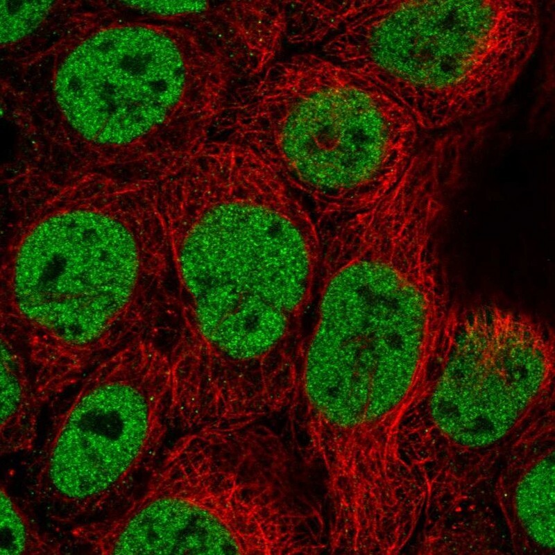
- Main image

- Experimental details
- Immunofluorescent staining of human cell line CACO-2 shows localization to nucleoplasm.
- Sample type
- Human
Supportive validation
- Submitted by
- Atlas Antibodies (provider)
- Enhanced method
- Orthogonal validation
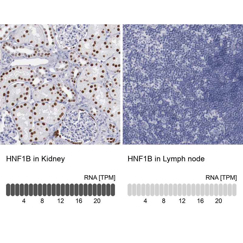
- Main image
- Experimental details
- Immunohistochemistry analysis in human kidney and lymph node tissues using HPA002083 antibody. Corresponding HNF1B RNA-seq data are presented for the same tissues.
- Sample type
- Human
- Protocol
- Protocol