 Explore
 Validate
 Learn
Learn Western blot
 Immunocytochemistry
Antibody data
- Antibody Data
- Antigen structure
- References [1]
- Comments [0]
- Validations
- Immunocytochemistry [1]
- Immunohistochemistry [1]
Submit
Validation data
Reference
Comment
Report error
- Product number
- AMAb90733 - Provider product page

- Provider
- Atlas Antibodies
- Proper citation
- Atlas Antibodies Cat#AMAb90733, RRID:AB_2665649
- Product name
- Anti-HNF1B
- Antibody type
- Monoclonal
- Description
- Monoclonal Antibody against Human HNF1B, Clone ID: CL0374, Gene description: HNF1 homeobox B, Alternative Gene Names: HNF1beta, LFB3, MODY5, TCF2, VHNF1, Validated applications: WB, IHC, ICC, Uniprot ID: P35680, Storage: Store at +4°C for short term storage. Long time storage is recommended at -20°C.
- Reactivity
- Human
- Host
- Mouse
- Conjugate
- Unconjugated
- Isotype
- IgG
- Antibody clone number
- CL0374
- Vial size
- 100 µl
- Concentration
- 1.0 mg/ml
- Storage
- Store at +4°C for short term storage. Long time storage is recommended at -20°C.
- Handling
- The antibody solution should be gently mixed before use.
Submitted references Napsin-A and AMACR are Superior to HNF-1β in Distinguishing Between Mesonephric Carcinomas and Clear Cell Carcinomas of the Gynecologic Tract
Pors J, Segura S, Cheng A, Ji J, Tessier-Cloutier B, Cochrane D, Fix D, Park K, Gilks B, Hoang L
Applied Immunohistochemistry & Molecular Morphology 2020;28(8):593-601
Applied Immunohistochemistry & Molecular Morphology 2020;28(8):593-601
No comments: Submit comment
Supportive validation
- Submitted by
- Atlas Antibodies (provider)
- Main image
- Experimental details
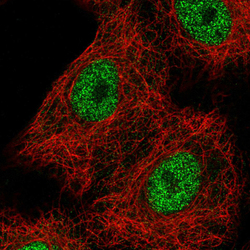
- Immunofluorescence staining in A549 cell line with Anti-HNF1B monoclonal antibody, showing spotty nuclear (without nucleoli) staining in green. Microtubule probes are visualized in red (where available).
- Sample type
- Human

Supportive validation
- Submitted by
- Atlas Antibodies (provider)
- Enhanced method
- Orthogonal validation
- Main image
- Experimental details
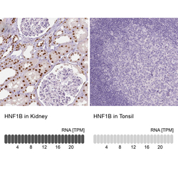
- Immunohistochemistry analysis in human kidney and tonsil tissues using AMAb90733 antibody. Corresponding HNF1B RNA-seq data are presented for the same tissues.
- Sample type
- Human
- Protocol
- Protocol
