 Explore
 Validate
 Learn
Learn Western blot
Antibody data
- Antibody Data
- Antigen structure
- References [0]
- Comments [0]
- Validations
- Western blot [1]
- Immunocytochemistry [3]
- Immunohistochemistry [2]
- Flow cytometry [2]
- Other assay [1]
Submit
Validation data
Reference
Comment
Report error
- Product number
- 700047 - Provider product page

- Provider
- Invitrogen Antibodies
- Product name
- Phospho-SMAD1/SMAD5 (Ser463, Ser465) Recombinant Rabbit Monoclonal Antibody (31H14L11)
- Antibody type
- Monoclonal
- Antigen
- Synthetic peptide
- Reactivity
- Human
- Host
- Rabbit
- Isotype
- IgG
- Antibody clone number
- 31H14L11
- Vial size
- 100 µg
- Concentration
- 0.5 mg/mL
- Storage
- Store at 4°C short term. For long term storage, store at -20°C, avoiding freeze/thaw cycles.
No comments: Submit comment
Supportive validation
- Submitted by
- Invitrogen Antibodies (provider)
- Main image
- Experimental details
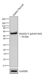
- Western blot analysis of SMAD1/5 (pS463/465) was performed by loading 20 µg of Rabbit Thyroid tissue lysate using Novex®NuPAGE®4-12% Bis-Tris gel (Product # NP0321BOX), XCell SureLock Electrophoresis System (Product # EI0002), Novex® Sharp Pre-Stained Protein Standard (Product # LC5800), and iBlot® Dry Blotting System (Product # IB21001). Proteins were transferred to a nitrocellulose membrane and blocked with 5% skim milk for 1 hour at room temperature. SMAD1/5 (pS463/465) was detected at ~58 kDa using SMAD1/5 (pS463/465) Recombinant Rabbit Monoclonal Antibody (Product # 700047) at 1-2 µg/mL in 2.5% skim milk at 4°C overnight on a rocking platform. Goat anti-Rabbit IgG - HRP Secondary Antibody (Product # G-21234) at 1:5000 dilution was used and chemiluminescent detection was performed using Pierce™ ECL Western blotting Substrate (Product # 32106).

Supportive validation
- Submitted by
- Invitrogen Antibodies (provider)
- Main image

- Experimental details
- Immunofluorescent analysis of Phospho-SMAD1/5 pSer463/465 in HeLa cells using a Phospho-SMAD1/5 pSer463/465 recombinant rabbit monoclonal antibody (Product # 700047) at a dilution of 2.5 µg/mL in the absence of peptide (top left) and presence of phosphopeptide used as immunogen (top right) or non-phosphopeptide (bottom left), followed by detection using an Alexa Fluor 488-conjugated goat anti-rabbit secondary antibody at a dilution of 1:1000. Actin was stained with Alexa Fluor 568 phalloidin (Product # A12380).

- Submitted by
- Invitrogen Antibodies (provider)
- Main image

- Experimental details
- Immunofluorescence analysis of SMAD1/5 (pS463/465) was done on 70% confluent log phase HeLa cells. The cells were fixed with 4% paraformaldehyde for 15 minutes, permeabilized with 0.25% Triton X-100 for 10 minutes, and blocked with 5% BSA for 1 hour at room temperature. The cells were labeled with SMAD1/5 (pS463/465) Recombinant Rabbit Monoclonal Antibody (Product # 700047) at 2 µg/mL in 1% BSA and incubated for 3 hours at room temperature and then labeled with Alexa Fluor 488 Goat anti-Rabbit IgG Secondary Antibody (Product # A-11008) at a dilution of 1:400 for 30 minutes at room temperature (Panel a: green). Nuclei (Panel b: blue) were stained with SlowFade® Gold Antifade Mountant DAPI (Product # S36938). F-actin (Panel c: red) was stained with Alexa Fluor 594 Phalloidin (Product # A12381). Panel d is a merged image showing nuclear localization. Panel e shows no primary antibody control. The images were captured at 20X magnification.

Supportive validation
- Submitted by
- Invitrogen Antibodies (provider)
- Main image
- Experimental details
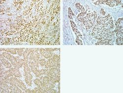
- Immunohistochemistry analysis of Phospho-SMAD1/5 pSer463/465 in formalin-fixed, paraffin-embedded human lung (top left), breast (top right) and thyroid carcimona (bottom) using a Phospho-SMAD1/5 pSer463/465 monoclonal antibody (Product # 700047) at a dilution of 5 µg/mL. Tissues were pretreated with EDTA and staining was visualized using DAB. Images were taken at a magnification of 20x. Results show nuclear staining in tumor cells.

- Submitted by
- Invitrogen Antibodies (provider)
- Main image
- Experimental details
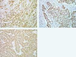
- Immunohistochemistry analysis of Phospho-SMAD1/5 pSer463/465 in formalin-fixed, paraffin-embedded human lung (top left), breast (top right) and thyroid carcimona (bottom) using a Phospho-SMAD1/5 pSer463/465 monoclonal antibody (Product # 700047) at a dilution of 5 µg/mL. Tissues were pretreated with EDTA and staining was visualized using DAB. Images were taken at a magnification of 20x. Results show nuclear staining in tumor cells.

Supportive validation
- Submitted by
- Invitrogen Antibodies (provider)
- Main image

- Experimental details
- Flow cytometry analysis of Smad1/5 [pS463/465] was done on serum starved HeLa cells. Cells were fixed with 70% ethanol for 10 minutes, permeabilized with 0.25% Triton™ X-100 for 20 minutes, and blocked with 5% BSA for 30 minutes at room temperature. Cells were labeled with ABfinity™ Smad1/5 [pS463/465] Recombinant Rabbit Monoclonal Antibody (700047, red histogram) or with rabbit isotype control (pink histogram) at 3-5 µg/million cells in 2.5% BSA. After incubation at room temperature for 2 hours, the cells were labeled with Alexa Fluor® 488 Goat Anti-Rabbit Secondary Antibody (A11008) at a dilution of 1:400 for 30 minutes at room temperature. The representative 10,000 cells were acquired and analyzed for each sample using an Attune® Acoustic Focusing Cytometer. The purple histogram represents unstained control cells and the green histogram represents no-primary-antibody control.

- Submitted by
- Invitrogen Antibodies (provider)
- Main image

- Experimental details
- Flow cytometry analysis of Phospho-SMAD1/5 pSer463/465 in Jurkat cells stimulated with BMP-4 (black) or unstimulated (gray) using a Phospho-SMAD1/5 pSer463/465 recombinant rabbit monoclonal antibody (Product # 700047) at a dilution of 0.5 µg. Cells were fixed and permeabilized using FIX & PERM (Product # GAS-004) reagent, and detection was performed using an Alexa Fluor 488 goat anti-rabbit IgG. Pre-incubation with the immunogenic peptide decreased the signal (red).

Supportive validation
- Submitted by
- Invitrogen Antibodies (provider)
- Main image

- Experimental details
- NULL
