 Explore
 Validate
 Learn
Learn Western blot
 Immunocytochemistry
Antibody data
- Antibody Data
- Antigen structure
- References [0]
- Comments [0]
- Validations
- Western blot [4]
- Immunocytochemistry [4]
- Immunohistochemistry [6]
Submit
Validation data
Reference
Comment
Report error
- Product number
- LS-C313035 - Provider product page

- Provider
- LSBio
- Product name
- p66 / SHC Antibody (aa424-440) LS-C313035
- Antibody type
- Polyclonal
- Description
- Immunogen affinity purified
- Reactivity
- Human, Mouse, Rat
- Host
- Rabbit
- Storage
- At -20°C for 1 year. After reconstitution, at 4°C for 1 month. It can also be aliquotted and stored frozen at -20°C for a longer time. Avoid freeze-thaw cycles.
No comments: Submit comment
Enhanced validation
- Submitted by
- LSBio (provider)
- Enhanced method
- Genetic validation
- Main image
- Experimental details
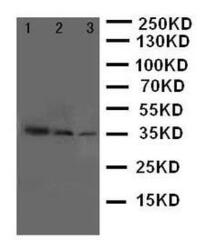
- WB of p66 / SHC antibody. Lane 1: Recombinant Human SHC1 Protein 10ng. Lane 2: Recombinant Human SHC1 Protein 5ng. Lane 3: Recombinant Human SHC1 Protein 2.5ng.

- Submitted by
- LSBio (provider)
- Enhanced method
- Genetic validation
- Main image

- Experimental details
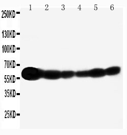
- WB of p66 / SHC antibody. Lane 1: Rat Brain Tissue Lysate. Lane 2: A549 Cell Lysate. Lane 3: A431 Cell Lysate. Lane 4: 293T Cell Lysate. Lane 5: HELA Cell Lysate. Lane 6: JURKAT Cell Lysate..
- Submitted by
- LSBio (provider)
- Enhanced method
- Genetic validation
- Main image

- Experimental details
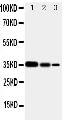
- Anti-SHC antibody, Western blottingRecombinant Protein Detection Source: E. coli derived -recombinant human SHC1, 35. 0KD(162aa tag+D424-P578)Lane 1: Recombinant Human SHC1 Proteins 10ng Lane 2: Recombinant Human SHC1 Proteins 5ng Lane 3: Recombinant Human SHC1 Proteins 2. 5ng
- Submitted by
- LSBio (provider)
- Enhanced method
- Genetic validation
- Main image
- Experimental details
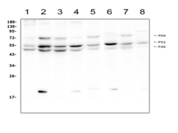
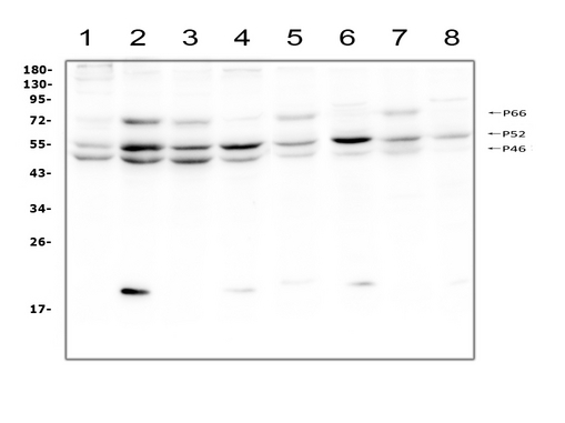
- Western blot analysis of SHC using anti-SHC antibody. Electrophoresis was performed on a 5-20% SDS-PAGE gel at 70V (Stacking gel) / 90V (Resolving gel) for 2-3 hours. The sample well of each lane was loaded with 50ug of sample under reducing conditions. Lane 1: human A431 whole cell lysate, Lane 2: human Hela whole cell lysate, Lane 3: human HepG2 whole cell lysate, Lane 4: human Jurkat whole cell lysate, Lane 5: rat C6 whole cell lysate, Lane 6: mouse thymus tissue lysate, Lane 7: mouse RAW246. 7 whole cell lysate, Lane 8: mouse NIH3T3 whole cell lysate. After Electrophoresis, proteins were transferred to a Nitrocellulose membrane at 150mA for 50-90 minutes. Blocked the membrane with 5% Non-fat Milk/ TBS for 1.5 hour at RT. The membrane was incubated with rabbit anti-SHC antigen affinity purified polyclonal antibody at 0.5 µg/mL overnight at 4°C, then washed with TBS-0.1% Tween 3 times with 5 minutes each and probed with a goat anti-rabbit IgG-HRP secondary antibody at a dilution of 1:10000 for 1.5 hour at RT. The signal is developed using an Enhanced Chemiluminescent detection (ECL) kit with Tanon 5200 system. Specific bands were detected for SHC at approximately 46, 52, 66KD. The expected band size for SHC are at 46, 52, 66KD.
Supportive validation
- Submitted by
- LSBio (provider)
- Enhanced method
- Genetic validation
- Main image

- Experimental details
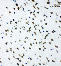
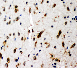
- p66 / SHC antibody. IHC(F): Rat Brain Tissue.

- Submitted by
- LSBio (provider)
- Enhanced method
- Genetic validation
- Main image

- Experimental details
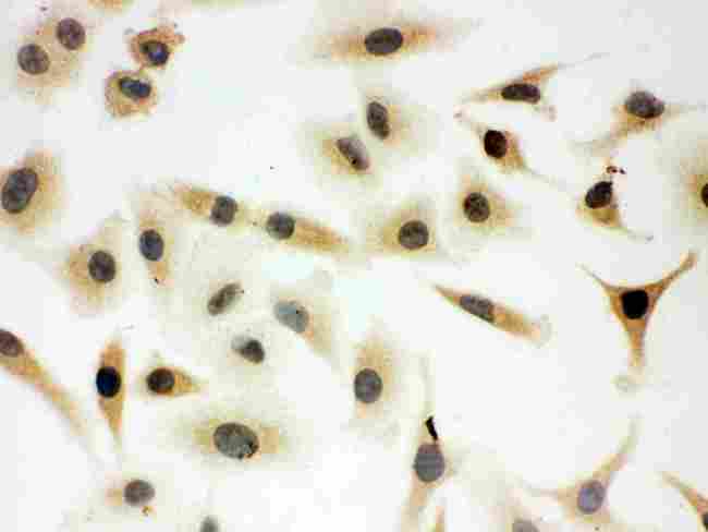
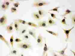
- Anti-SHC antibody, ICCICC: A549 Cell
- Submitted by
- LSBio (provider)
- Main image
- Experimental details
- p66 / SHC antibody. IHC(F): Rat Brain Tissue.
- Submitted by
- LSBio (provider)
- Main image
- Experimental details
- Anti-SHC antibody, ICCICC: A549 Cell

Supportive validation
- Submitted by
- LSBio (provider)
- Enhanced method
- Genetic validation
- Main image
- Experimental details
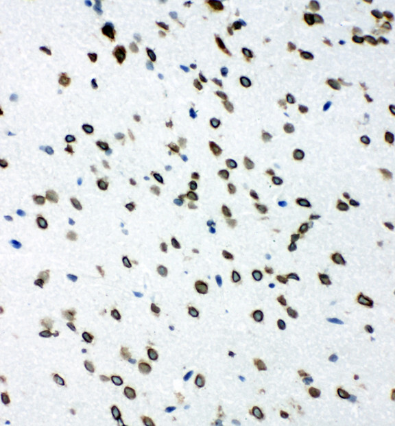
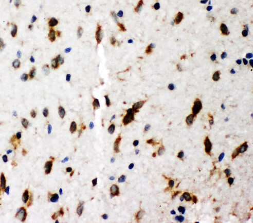
- p66 / SHC antibody. IHC(P): Rat Brain Tissue.
- Submitted by
- LSBio (provider)
- Enhanced method
- Genetic validation
- Main image

- Experimental details
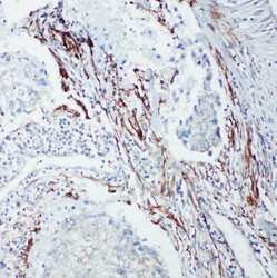
- p66 / SHC antibody. IHC(P): Human Lung Cancer Tissue.

- Submitted by
- LSBio (provider)
- Enhanced method
- Genetic validation
- Main image

- Experimental details
- p66 / SHC antibody. IHC(F): Rat Intestine Tissue.

- Submitted by
- LSBio (provider)
- Enhanced method
- Genetic validation
- Main image

- Experimental details
- p66 / SHC antibody. IHC(P): Rat Brain Tissue.

- Submitted by
- LSBio (provider)
- Enhanced method
- Genetic validation
- Main image
- Experimental details
- p66 / SHC antibody. IHC(P): Human Lung Cancer Tissue.

- Submitted by
- LSBio (provider)
- Enhanced method
- Genetic validation
- Main image

- Experimental details
- p66 / SHC antibody. IHC(F): Rat Intestine Tissue.
