 Explore
 Validate
 Learn
Learn Western blot
Antibody data
- Antibody Data
- Antigen structure
- References [0]
- Comments [0]
- Validations
- Western blot [2]
- Immunohistochemistry [1]
Submit
Validation data
Reference
Comment
Report error
- Product number
- GTX24890 - Provider product page

- Provider
- GeneTex
- Proper citation
- GeneTex Cat#GTX24890, RRID:AB_380458
- Product name
- SHC1 (phospho Tyr239/Tyr240) antibody
- Antibody type
- Polyclonal
- Reactivity
- Human, Mouse, Rat
- Host
- Rabbit
No comments: Submit comment
Supportive validation
- Submitted by
- GeneTex (provider)
- Main image

- Experimental details
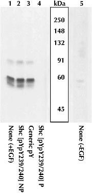
- Extracts prepared from A431 and stimulated with EGF (1-4) or left unstimulated with EGF (5) were resolved by SDS-PAGE on a 10% Tris-glycine gel, and transferred to PVDF. Membranes were blocked with a 5% BSA-TBST buffer overnight and then incubated with GTX24890
- Submitted by
- GeneTex (provider)
- Main image
- Experimental details
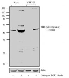
- Western blot analysis was performed on whole cell extracts (20 ug lysate) of A431 (Lane 1), A431 treated for 10 minutes with 200 ng/ml of EGF (Lane 2), NIH/3T3 (Lane 3) and NIH/3T3 treated for 10 minutes with 200 ng/ml of EGF (lane 4). The blots were probed with Anti-SHC [pY239/pY240] Rabbit Polyclonal Antibody and detected by chemiluminescence Goat anti-Rabbit IgG (H+L) Superclonal? Secondary Antibody, HRP conjugate. A 51 kDa band corresponding to SHC [pY239/pY240] was observed across the EGF treated cell lines tested. Known quantity of protein samples were electrophoresed using Novex? NuPAGE? 12 % Bis-Tris gel, XCell SureLock? Electrophoresis System and Novex? Sharp Pre-Stained Protein Standard. Resolved proteins were then transferred onto a nitrocellulose membrane with iBlot? 2 Dry Blotting System. The membrane was probed with the relevant primary and secondary Antibody following blocking with 5 % skimmed milk. Chemiluminescent detection was performed using Novex? ECL Chemiluminescent Substrate Reagent Kit.

Supportive validation
- Submitted by
- GeneTex (provider)
- Main image
- Experimental details
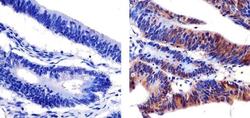
- Immunohistochemistry analysis of SHC [PYPY239/240] showing staining in the cytoplasm and nucleus of paraffin-embedded human colon carcinoma (right) compared to a negative control without primary antibody (left). To expose target proteins, antigen retrieval was performed using 10mM sodium citrate (pH 6.0), microwaved for 8-15 min. Following antigen retrieval, tissues were blocked in 3% H2O2-methanol for 15 min at room temperature, washed with ddH2O and PBS, and then probed with a SHC [PYPY239/240] Rabbit Polyclonal Antibody diluted in 3% BSA-PBS at a dilution of 1:20 overnight at 4¢XC in a humidified chamber. Tissues were washed extensively in PBST and detection was performed using an HRP-conjugated secondary antibody followed by colorimetric detection using a DAB kit. Tissues were counterstained with hematoxylin and dehydrated with ethanol and xylene to prep for mounting.
