 Explore
 Validate
 Learn
Learn Western blot
 Immunocytochemistry
Antibody data
- Antibody Data
- Antigen structure
- References [2]
- Comments [0]
- Validations
- Immunocytochemistry [4]
- Immunohistochemistry [1]
- Other assay [3]
Submit
Validation data
Reference
Comment
Report error
- Product number
- MA5-16147 - Provider product page

- Provider
- Invitrogen Antibodies
- Product name
- DUX4 Monoclonal Antibody (P4H2)
- Antibody type
- Monoclonal
- Antigen
- Synthetic peptide
- Reactivity
- Human
- Host
- Mouse
- Isotype
- IgG
- Antibody clone number
- P4H2
- Vial size
- 100 μL
- Concentration
- 1 mg/mL
- Storage
- Store at 4°C short term. For long term storage, store at -20°C, avoiding freeze/thaw cycles.
Submitted references Superficial sarcomas with CIC rearrangement are aggressive neoplasms: A series of eight cases.
DUX4 Immunohistochemistry Is a Highly Sensitive and Specific Marker for CIC-DUX4 Fusion-positive Round Cell Tumor.
Ko JS, Marusic Z, Azzato EM, Farkas DH, Van Arnam J, Seiwerth S, Fritchie K, Patel RM, Rubin BP, Billings SD
Journal of cutaneous pathology 2020 Jun;47(6):509-516
Journal of cutaneous pathology 2020 Jun;47(6):509-516
DUX4 Immunohistochemistry Is a Highly Sensitive and Specific Marker for CIC-DUX4 Fusion-positive Round Cell Tumor.
Siegele B, Roberts J, Black JO, Rudzinski E, Vargas SO, Galambos C
The American journal of surgical pathology 2017 Mar;41(3):423-429
The American journal of surgical pathology 2017 Mar;41(3):423-429
No comments: Submit comment
Supportive validation
- Submitted by
- Invitrogen Antibodies (provider)
- Main image
- Experimental details
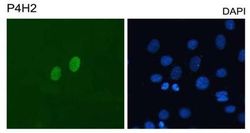
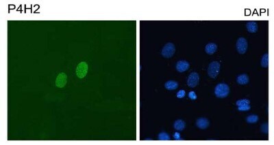
- Immunofluorescent analysis of DUX4 using a monoclonal antibody (Product # MA5-16147).
- Submitted by
- Invitrogen Antibodies (provider)
- Main image
- Experimental details
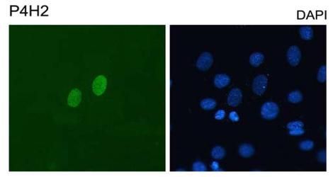
- Immunocytochemistry analysis of DUX4 in C2C12 mouse myoblasts transfected with pCS2-DUX4. Samples were incubated in DUX4 monoclonal antibody (Product # MA5-16147). Cells were counterstained with DAPI for nuclei.
- Submitted by
- Invitrogen Antibodies (provider)
- Main image

- Experimental details
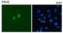
- Immunocytochemistry analysis of DUX4 in C2C12 mouse myoblasts transfected with pCS2-DUX4. Samples were incubated in DUX4 monoclonal antibody (Product # MA5-16147). Cells were counterstained with DAPI for nuclei.
- Submitted by
- Invitrogen Antibodies (provider)
- Main image

- Experimental details
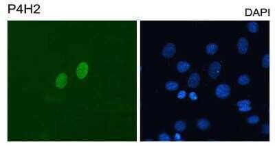
- Immunocytochemistry analysis of DUX4 in C2C12 mouse myoblasts transfected with pCS2-DUX4. Samples were incubated in DUX4 monoclonal antibody (Product # MA5-16147). Cells were counterstained with DAPI for nuclei.

Supportive validation
- Submitted by
- Invitrogen Antibodies (provider)
- Main image

- Experimental details
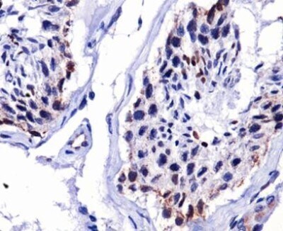
- Immunohistochemical analysis of DUX4 in human testis. Samples were incubated in DUX4 monoclonal antibody (Product # MA5-16147).
Supportive validation
- Submitted by
- Invitrogen Antibodies (provider)
- Main image

- Experimental details
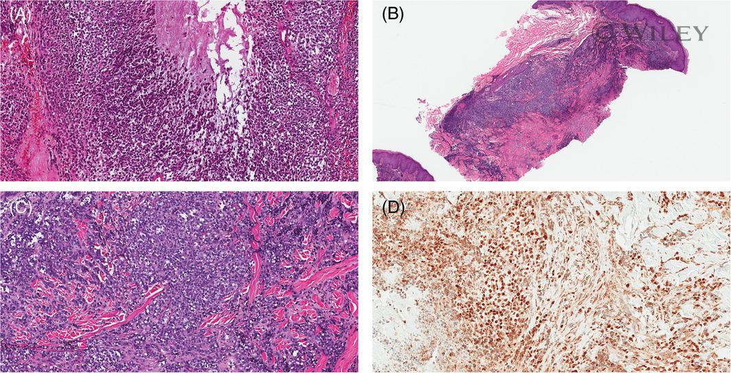
- A, Case 6. Occasional microcystic and corded architecture (hematoxylin and eosin [H&E], x200) noted. B and C. Case 3, Foci of rhabdoid and plasmacytoid features (H&E, x40 and x200). D, A representative image of DUX4 immunohistochemical staining is from case 7 (x200)
- Submitted by
- Invitrogen Antibodies (provider)
- Main image
- Experimental details
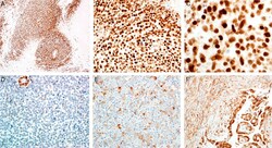
- FIGURE 2. Positive DUX4 monoclonal antibody staining demonstrating tumor cell-specific strong, crisp nuclear staining in representative sections of a CIC-DUX4 fusion-positive round cell tumor at low-power magnification (DUX4) (A) and at high-power magnifications (DUX4) (B and C). D, Negative DUX4 staining demonstrating complete lack of tumor cell staining in a Ewing sarcoma (DUX4). E, Negative DUX4 staining demonstrating absent nuclear staining and patchy mild cytoplasmic staining in a malignant rhabdoid tumor (DUX4). F, Negative DUX4 staining demonstrating absent nuclear staining and diffuse moderate, with focal strong, cytoplasmic staining in a synovial sarcoma, biphasic type (DUX4).

- Submitted by
- Invitrogen Antibodies (provider)
- Main image

- Experimental details
- FIGURE 3. A-E, S1-S5, respectively. Positive DUX4 monoclonal antibody staining in 5 separate CIC-DUX4 fusion-positive round cell tumors (H&E, DUX4).
