 Explore
 Validate
 Learn
Learn Western blot
Antibody data
- Antibody Data
- Antigen structure
- References [0]
- Comments [0]
- Validations
- Western blot [4]
- Immunohistochemistry [3]
Submit
Validation data
Reference
Comment
Report error
- Product number
- ABIN2506494 - Provider product page

- Provider
- antibodies-online
- Product name
- anti-Transcription Elongation Factor, Mitochondrial (TEFM) (N-Term) antibody
- Antibody type
- Polyclonal
- Antigen
- The immunogen for Anti-TEFM antibody is: synthetic peptide directed towards the N-terminal region of Human TEFM
- Description
- Affinity Purified
- Reactivity
- Human
- Host
- Rabbit
- Antigen sequence
RSSLYWALHN FCCRKKSTTP KKITPNVTFC DE
NAKEPENA LDKLFSSEQQ- Epitope
- N-Term
- Vial size
- 50 μg
- Storage
- Add 50 μl of distilled water. Final Anti-TEFM antibody concentration is 1 mg/mL in PBS buffer with 2 % sucrose. For longer periods of storage, store at -20°C. Avoid repeat freeze-thaw cycles.
- Handling
- Avoid repeat freeze-thaw cycles.
No comments: Submit comment
Supportive validation
- Submitted by
- antibodies-online (provider)
- Main image
- Experimental details
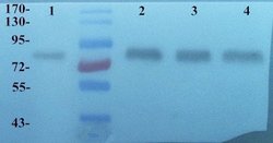
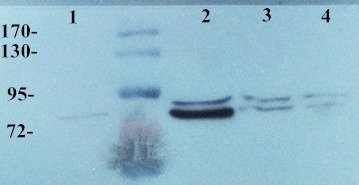
- Western blot analysis of Mouse spleen (Lane 1), Rat lung (Lane 2), Rat spleen (Lane 3), Rat heart (Land 4) using MMP9 antibody (Dilution at 2 ug/ml)
- Submitted by
- antibodies-online (provider)
- Main image
- Experimental details
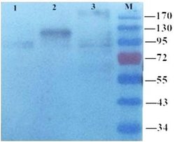
- Western blot analysis of rat brain (Land 1), kidney (Land 2) and lung (Land 3) using MMP9 antibody (dilution of primary antibody at 1:100)
- Submitted by
- antibodies-online (provider)
- Main image

- Experimental details
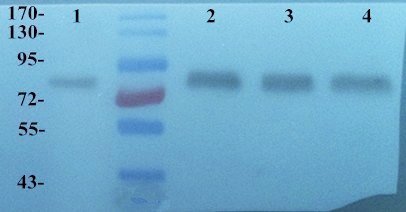
- WB analysis of Mouse spleen (Lane 1), Rat lung (Lane 2), Rat spleen (Lane 3), Rat heart (Lane 4) using anti MMP9 (primary antibody dilution at 2 ug/ml)
- Submitted by
- antibodies-online (provider)
- Main image

- Experimental details
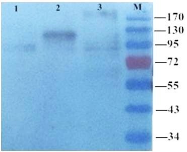
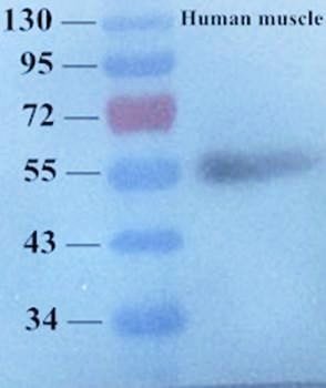
- Western blot analysis of human muscle tissue using MMP9 antibody (primary antibody at 1:100)
Supportive validation
- Submitted by
- antibodies-online (provider)
- Main image
- Experimental details
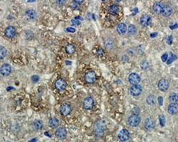
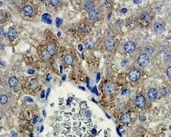
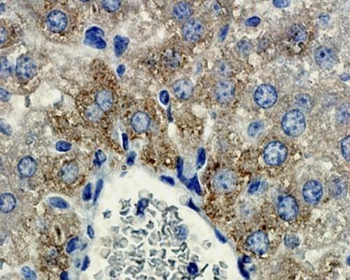
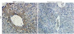
- IHC-P staining of mouse liver tissue using anti-MMP9 (dilution at 1:200)
- Submitted by
- antibodies-online (provider)
- Main image
- Experimental details
- IHC-P image of mouse liver tissue using MMP9 antibody (dilution of primary antibody at 1:200)
- Submitted by
- antibodies-online (provider)
- Main image
- Experimental details
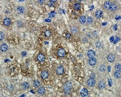
- IHC-P staining of mouse liver tissue using MMP9 antibody (dilution at 1:200)
