Explore
 Validate
 Learn
LearnMA5-32771
antibody from Invitrogen Antibodies
Targeting: TXNIP
ARRDC6, EST01027, HHCPA78, THIF, VDUP1
 Western blot
 Immunocytochemistry
Antibody data
- Antibody Data
- Antigen structure
- References [1]
- Comments [0]
- Validations
- Immunocytochemistry [6]
- Immunohistochemistry [4]
- Flow cytometry [2]
- Other assay [1]
Submit
Validation data
Reference
Comment
Report error
- Product number
- MA5-32771 - Provider product page

- Provider
- Invitrogen Antibodies
- Product name
- TXNIP Recombinant Rabbit Monoclonal Antibody (JM60-35)
- Antibody type
- Monoclonal
- Antigen
- Synthetic peptide
- Description
- Recombinant rabbit monoclonal antibodies are produced using in vitro expression systems. The expression systems are developed by cloning in the specific antibody DNA sequences from immunoreactive rabbits. Then, individual clones are screened to select the best candidates for production. The advantages of using recombinant rabbit monoclonal antibodies include: better specificity and sensitivity, lot-to-lot consistency, animal origin-free formulations, and broader immunoreactivity to diverse targets due to larger rabbit immune repertoire.
- Reactivity
- Human, Mouse, Rat
- Host
- Rabbit
- Isotype
- IgG
- Antibody clone number
- JM60-35
- Vial size
- 100 μL
- Concentration
- 1 mg/mL
- Storage
- Store at 4°C short term. For long term storage, store at -20°C, avoiding freeze/thaw cycles.
Submitted references TXNIP Participated in NLRP3-Mediated Inflammation in a Rat Model of Cervical Spondylotic Myelopathy.
Liu P, Li X, Liu J, Zhang H, You Z, Zhang J
Journal of inflammation research 2022;15:4547-4559
Journal of inflammation research 2022;15:4547-4559
No comments: Submit comment
Supportive validation
- Submitted by
- Invitrogen Antibodies (provider)
- Main image

- Experimental details
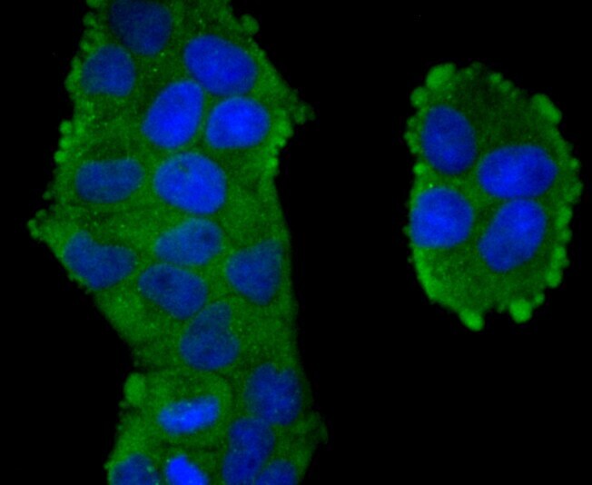
- Immunocytochemical analysis of TXNIP in Hela cells using a TXNIP Monoclonal antibody (Product # MA5-32771) as seen in green. The nuclear counter stain is DAPI (blue). Cells were fixed in paraformaldehyde, permeabilised with 0.25% Triton X100/PBS.
- Submitted by
- Invitrogen Antibodies (provider)
- Main image

- Experimental details
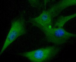
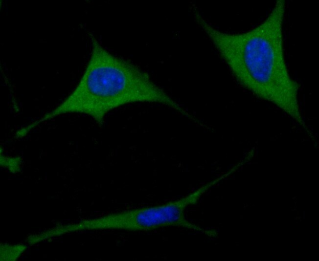
- Immunocytochemical analysis of TXNIP in NIH-3T3 cells using a TXNIP Monoclonal antibody (Product # MA5-32771) as seen in green. The nuclear counter stain is DAPI (blue). Cells were fixed in paraformaldehyde, permeabilised with 0.25% Triton X100/PBS.

- Submitted by
- Invitrogen Antibodies (provider)
- Main image

- Experimental details
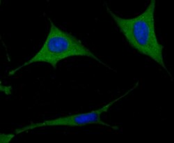
- Immunocytochemical analysis of TXNIP in SH-SY5Y cells using a TXNIP Monoclonal antibody (Product # MA5-32771) as seen in green. The nuclear counter stain is DAPI (blue). Cells were fixed in paraformaldehyde, permeabilised with 0.25% Triton X100/PBS.

- Submitted by
- Invitrogen Antibodies (provider)
- Main image

- Experimental details
- Immunocytochemical analysis of TXNIP in Hela cells using a TXNIP Monoclonal antibody (Product # MA5-32771) as seen in green. The nuclear counter stain is DAPI (blue). Cells were fixed in paraformaldehyde, permeabilised with 0.25% Triton X100/PBS.

- Submitted by
- Invitrogen Antibodies (provider)
- Main image
- Experimental details
- Immunocytochemical analysis of TXNIP in NIH-3T3 cells using a TXNIP Monoclonal antibody (Product # MA5-32771) as seen in green. The nuclear counter stain is DAPI (blue). Cells were fixed in paraformaldehyde, permeabilised with 0.25% Triton X100/PBS.

- Submitted by
- Invitrogen Antibodies (provider)
- Main image
- Experimental details
- Immunocytochemical analysis of TXNIP in SH-SY5Y cells using a TXNIP Monoclonal antibody (Product # MA5-32771) as seen in green. The nuclear counter stain is DAPI (blue). Cells were fixed in paraformaldehyde, permeabilised with 0.25% Triton X100/PBS.
Supportive validation
- Submitted by
- Invitrogen Antibodies (provider)
- Main image
- Experimental details
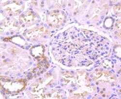
- Immunohistochemical analysis of TXNIP of paraffin-embedded Human kidney tissue using a TXNIP Monoclonal antibody (Product #MA5-32771). Counter stained with hematoxylin.

- Submitted by
- Invitrogen Antibodies (provider)
- Main image
- Experimental details
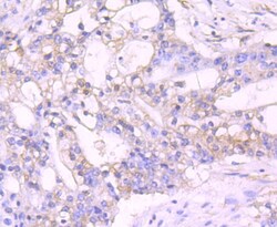
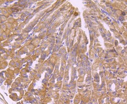
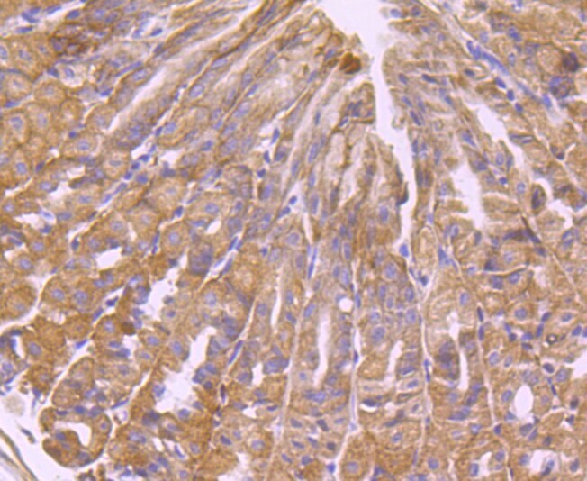
- Immunohistochemical analysis of TXNIP of paraffin-embedded Human stomach cancer tissue using a TXNIP Monoclonal antibody (Product #MA5-32771). Counter stained with hematoxylin.

- Submitted by
- Invitrogen Antibodies (provider)
- Main image

- Experimental details
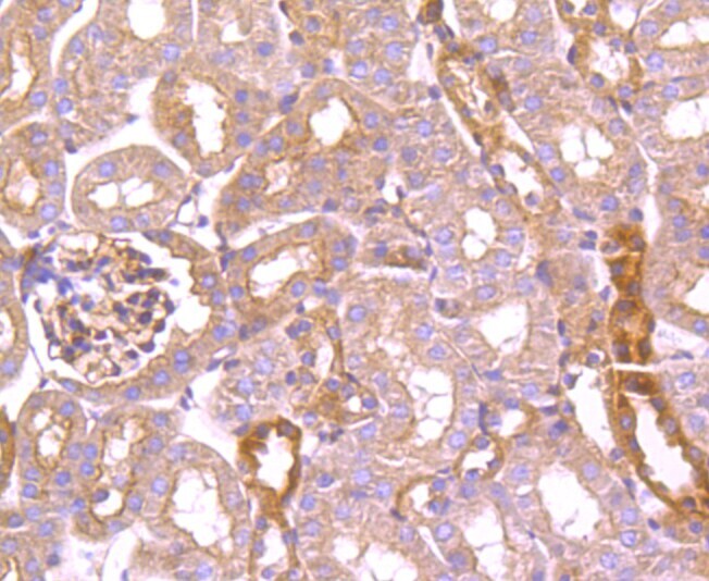
- Immunohistochemical analysis of TXNIP of paraffin-embedded Mouse kidney tissue using a TXNIP Monoclonal antibody (Product #MA5-32771). Counter stained with hematoxylin.
- Submitted by
- Invitrogen Antibodies (provider)
- Main image
- Experimental details
- Immunohistochemical analysis of TXNIP of paraffin-embedded Mouse stomach tissue using a TXNIP Monoclonal antibody (Product #MA5-32771). Counter stained with hematoxylin.
Supportive validation
- Submitted by
- Invitrogen Antibodies (provider)
- Main image
- Experimental details
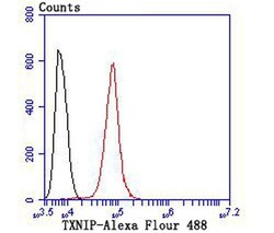
- Flow Cytometric analysis of TXNIP in Hela cells using a TXNIP Monoclonal Antibody (Product # MA5-32771) at a dilution of 1:100, as seen in red compared with an unlabelled control (cells without incubation with primary antibody; black).
- Submitted by
- Invitrogen Antibodies (provider)
- Main image

- Experimental details
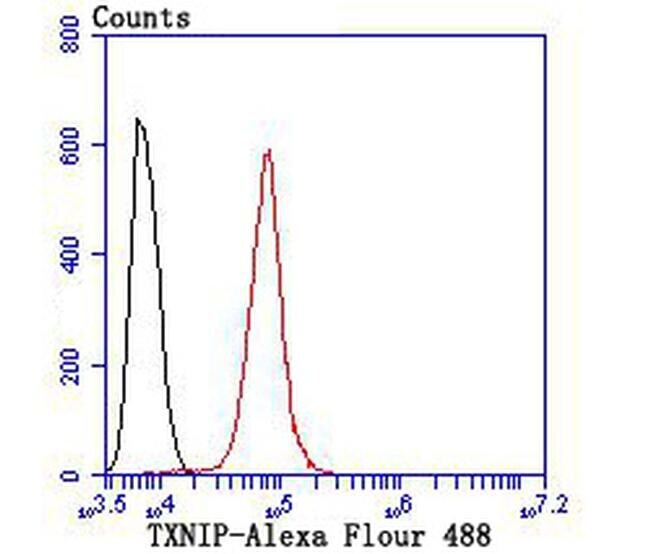
- Flow Cytometric analysis of TXNIP in Hela cells using a TXNIP Monoclonal Antibody (Product # MA5-32771) at a dilution of 1:100, as seen in red compared with an unlabelled control (cells without incubation with primary antibody; black).

Supportive validation
- Submitted by
- Invitrogen Antibodies (provider)
- Main image
- Experimental details
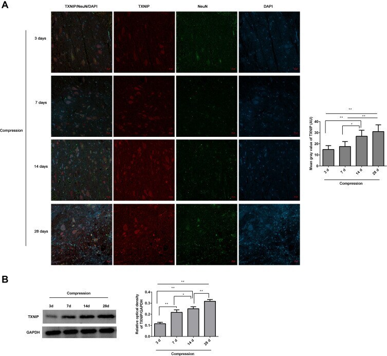
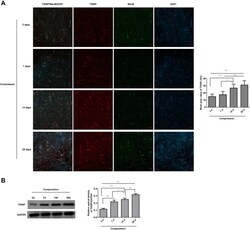
- The expression of TNXIP in the lesioned spinal cord following compression was observed. ( A ) The TNXIP/NeuN expression in the anterior horn of lesioned spinal cords was analyzed by immunofluorescence. The mean fluorescence intensities of TXNIP were analyzed by Image J software, and represented as mean gray values. ( B ) The TXNIP expression in the lesioned spinal cords was analyzed by Western blot. The gray values of protein bands were analyzed by Image J software, and protein expression was normalized to GAPDH. *P