Explore
 Validate
 Learn
Learn Western blot
 Immunocytochemistry
 Immunohistochemistry
Antibody data
- Antibody Data
- Antigen structure
- References [6]
- Comments [0]
- Validations
- Western blot [1]
- Immunocytochemistry [1]
Submit
Validation data
Reference
Comment
Report error
- Product number
- HPA001330 - Provider product page

- Provider
- Atlas Antibodies
- Proper citation
- Atlas Antibodies Cat#HPA001330, RRID:AB_1080167
- Product name
- Anti-STX4
- Antibody type
- Polyclonal
- Description
- Polyclonal Antibody against Human STX4, Gene description: syntaxin 4, Alternative Gene Names: p35-2, STX4A, Validated applications: ICC, IHC, WB, Uniprot ID: Q12846, Storage: Store at +4°C for short term storage. Long time storage is recommended at -20°C.
- Reactivity
- Human
- Host
- Rabbit
- Conjugate
- Unconjugated
- Isotype
- IgG
- Vial size
- 100 µl
- Concentration
- 0.3 mg/ml
- Storage
- Store at +4°C for short term storage. Long time storage is recommended at -20°C.
- Handling
- The antibody solution should be gently mixed before use.
Submitted references Morphological and histochemical characterization of the secretory epithelium in the canine lacrimal gland.
Canine Salivary Glands: Analysis of Rab and SNARE Protein Expression and SNARE Complex Formation With Diverse Tissue Properties.
Ultrastructure and immunohistochemical characterization of proteins concerned with the secretory machinery in goat ceruminous glands.
Tctex1d2 Is a Negative Regulator of GLUT4 Translocation and Glucose Uptake
Quantitative Proteomics Reveals That Only a Subset of the Endoplasmic Reticulum Contributes to the Phagosome
Protein expression of PKCZ (Protein Kinase C Zeta), Munc18c, and Syntaxin-4 in the insulin pathway in endometria of patients with polycystic ovary syndrome (PCOS)
Yasui T, Miyata K, Nakatsuka C, Tsukise A, Gomi H
European journal of histochemistry : EJH 2021 Nov 2;65(4)
European journal of histochemistry : EJH 2021 Nov 2;65(4)
Canine Salivary Glands: Analysis of Rab and SNARE Protein Expression and SNARE Complex Formation With Diverse Tissue Properties.
Gomi H, Osawa H, Uno R, Yasui T, Hosaka M, Torii S, Tsukise A
The journal of histochemistry and cytochemistry : official journal of the Histochemistry Society 2017 Nov;65(11):637-653
The journal of histochemistry and cytochemistry : official journal of the Histochemistry Society 2017 Nov;65(11):637-653
Ultrastructure and immunohistochemical characterization of proteins concerned with the secretory machinery in goat ceruminous glands.
Yasui T, Gomi H, Kitahara T, Tsukise A
European journal of histochemistry : EJH 2017 Aug 8;61(3):2828
European journal of histochemistry : EJH 2017 Aug 8;61(3):2828
Tctex1d2 Is a Negative Regulator of GLUT4 Translocation and Glucose Uptake
Shimoda Y, Okada S, Yamada E, Pessin J, Yamada M
Endocrinology 2015;156(10):3548-3558
Endocrinology 2015;156(10):3548-3558
Quantitative Proteomics Reveals That Only a Subset of the Endoplasmic Reticulum Contributes to the Phagosome
Campbell-Valois F, Trost M, Chemali M, Dill B, Laplante A, Duclos S, Sadeghi S, Rondeau C, Morrow I, Bell C, Gagnon E, Hatsuzawa K, Thibault P, Desjardins M
Molecular & Cellular Proteomics 2012;11(7):M111.016378-1-M111.016378-13
Molecular & Cellular Proteomics 2012;11(7):M111.016378-1-M111.016378-13
Protein expression of PKCZ (Protein Kinase C Zeta), Munc18c, and Syntaxin-4 in the insulin pathway in endometria of patients with polycystic ovary syndrome (PCOS)
Rivero R, Garin C, Ormazabal P, Silva A, Carvajal R, Gabler F, Romero C, Vega M
Reproductive Biology and Endocrinology 2012;10(1)
Reproductive Biology and Endocrinology 2012;10(1)
No comments: Submit comment
Enhanced validation
- Submitted by
- Atlas Antibodies (provider)
- Enhanced method
- Genetic validation
- Main image
- Experimental details
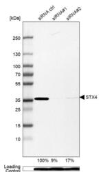
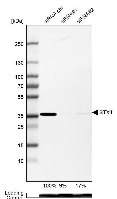
- Western blot analysis in A-549 cells transfected with control siRNA, target specific siRNA probe #1 and #2, using Anti-STX4 antibody. Remaining relative intensity is presented. Loading control: Anti-PPIB.
- Sample type
- Human
- Protocol
- Protocol
Supportive validation
- Submitted by
- Atlas Antibodies (provider)
- Main image

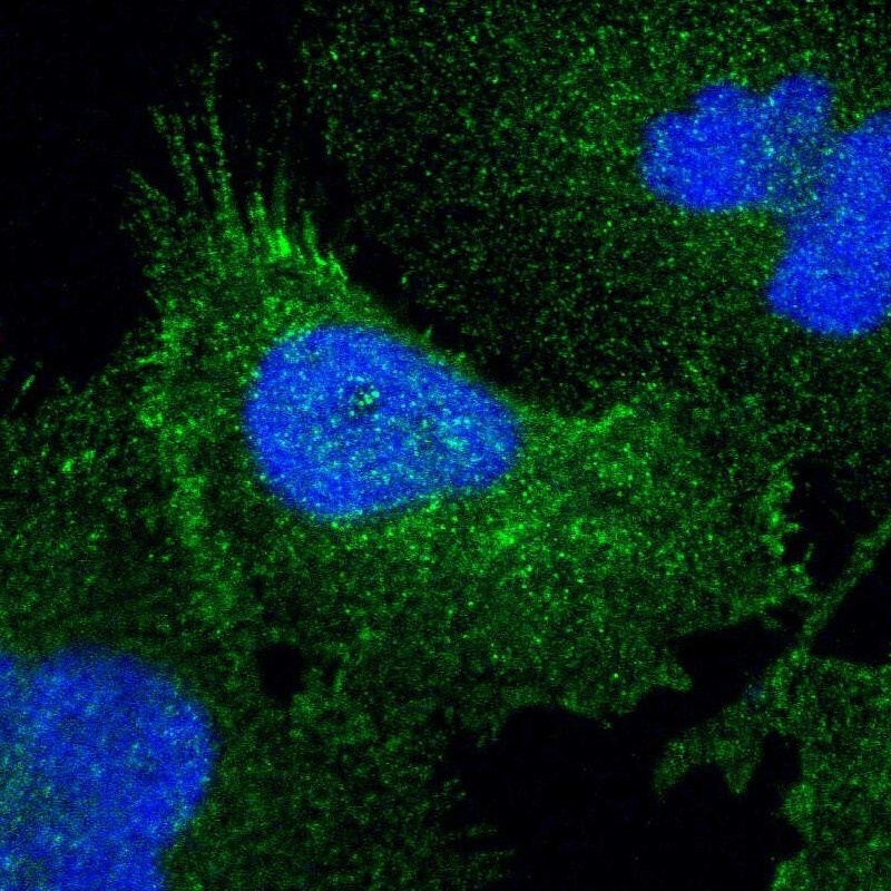
- Experimental details
- Immunofluorescent staining of human cell line U-251 MG shows localization to plasma membrane.
- Sample type
- Human