Explore
 Validate
 Learn
Learn Western blot
Antibody data
- Antibody Data
- Antigen structure
- References [2]
- Comments [0]
- Validations
- Western blot [6]
- Immunocytochemistry [1]
- Immunoprecipitation [1]
- Immunohistochemistry [3]
Submit
Validation data
Reference
Comment
Report error
- Product number
- GTX100748 - Provider product page

- Provider
- GeneTex
- Proper citation
- GeneTex Cat#GTX100748, RRID:AB_1952121
- Product name
- Syk antibody [N2C2], Internal
- Antibody type
- Polyclonal
- Reactivity
- Human, Mouse, Rat
- Host
- Rabbit
Submitted references Tetrandrine enhances the ubiquitination and degradation of Syk through an AhR-c-src-c-Cbl pathway and consequently inhibits osteoclastogenesis and bone destruction in arthritis.
Tetrandrine attenuates the bone erosion in collagen-induced arthritis rats by inhibiting osteoclastogenesis via spleen tyrosine kinase.
Jia Y, Tao Y, Lv C, Xia Y, Wei Z, Dai Y
Cell death & disease 2019 Jan 15;10(2):38
Cell death & disease 2019 Jan 15;10(2):38
Tetrandrine attenuates the bone erosion in collagen-induced arthritis rats by inhibiting osteoclastogenesis via spleen tyrosine kinase.
Jia Y, Miao Y, Yue M, Shu M, Wei Z, Dai Y
FASEB journal : official publication of the Federation of American Societies for Experimental Biology 2018 Jun;32(6):3398-3410
FASEB journal : official publication of the Federation of American Societies for Experimental Biology 2018 Jun;32(6):3398-3410
No comments: Submit comment
Supportive validation
- Submitted by
- GeneTex (provider)
- Main image
- Experimental details
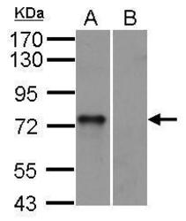
- SYK antibody [N2C2], Internal detects SYK protein by western blot analysis.A. 30 ?g mouse BMDM (bone marrow-derived macrophage) cellsB. 30 ?g mouse Syk null cells10% SDS-PAGESYK antibody [N2C2], Internal (GTX100748) dilution: 1:1000 The HRP-conjugated anti-rabbit IgG antibody (GTX213110-01) was used to detect the primary antibody.

- Submitted by
- GeneTex (provider)
- Main image

- Experimental details
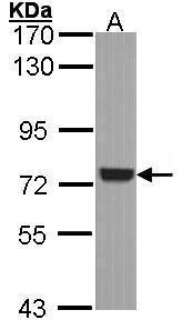
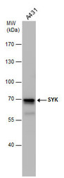
- Sample (30 ug of whole cell lysate) A: A431 (GTX27909) 7.5% SDS PAGE GTX100748 diluted at 1:1000
- Validation comment
- WB
- Submitted by
- GeneTex (provider)
- Main image

- Experimental details
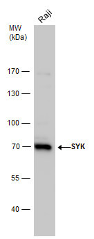
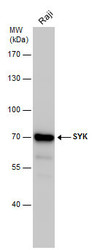
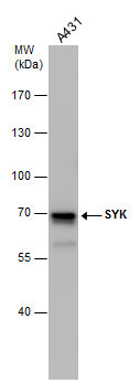
- SYK antibody detects SYK protein by western blot analysis. Whole cell extracts (30 ?g) was separated by 7.5% SDS-PAGE, and the membrane was blotted with SYK antibody (GTX100748) diluted by 1:2000.
- Validation comment
- WB
- Submitted by
- GeneTex (provider)
- Main image
- Experimental details
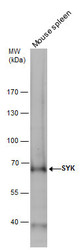
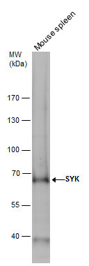
- SYK antibody detects SYK protein by western blot analysis. Mouse tissue extracts (50 ?g) was separated by 7.5% SDS-PAGE, and the membrane was blotted with SYK antibody (GTX100748) diluted by 1:1000. The HRP-conjugated anti-rabbit IgG antibody (GTX213110-01) was used to detect the primary antibody.
- Submitted by
- GeneTex (provider)
- Main image
- Experimental details
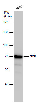
- SYK antibody detects SYK protein by western blot analysis. Whole cell extracts (30 ?g) was separated by 7.5% SDS-PAGE, and the membrane was blotted with SYK antibody (GTX100748) at a dilution of 1:2000. The HRP-conjugated anti-rabbit IgG antibody (GTX213110-01) was used to detect the primary antibody.
- Submitted by
- GeneTex (provider)
- Main image
- Experimental details
- SYK antibody detects SYK protein by western blot analysis. Whole cell extracts (30 ?g) was separated by 7.5% SDS-PAGE, and the membrane was blotted with SYK antibody (GTX100748) at a dilution of 1:2000. The HRP-conjugated anti-rabbit IgG antibody (GTX213110-01) was used to detect the primary antibody.
Supportive validation
- Submitted by
- GeneTex (provider)
- Main image
- Experimental details
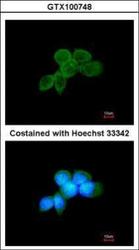
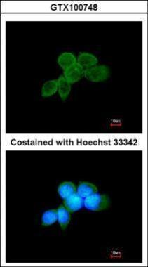
- Immunofluorescence analysis of methanol-fixed A431, using SYK(GTX100748) antibody at 1:200 dilution.
Supportive validation
- Submitted by
- GeneTex (provider)
- Main image
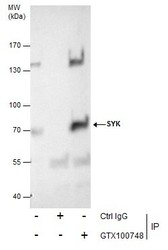
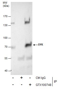
- Experimental details
- Immunoprecipitation of SYK protein from A431 whole cell extracts using 5 £gg of SYK antibody [N2C2] (GTX100748).Western blot analysis was performed using SYK antibody [N2C2] (GTX100748).EasyBlot anti-Rabbit IgG (GTX221666-01) was used as a secondary reagent.
Supportive validation
- Submitted by
- GeneTex (provider)
- Main image
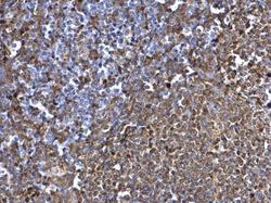
- Experimental details
- SYK antibody [N2C2], Internal detects SYK protein at cytosol on rat spleen by immunohistochemical analysis. Sample: Paraffin-embedded rat spleen. SYK antibody [N2C2], Internal (GTX100748) dilution: 1:500.

- Submitted by
- GeneTex (provider)
- Main image

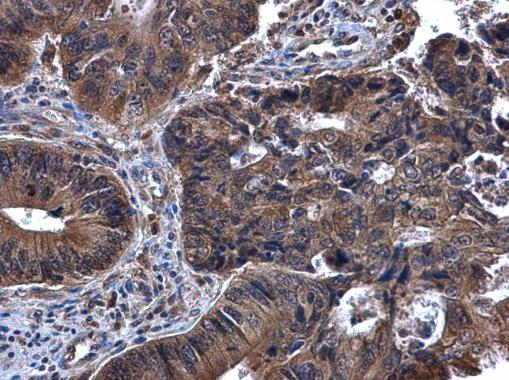
- Experimental details
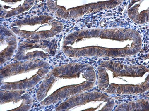
- SYK antibody [N2C2], Internal detects SYK protein at cytoplasm in human cervical cancer by immunohistochemical analysis. Sample: Paraffin-embedded human cervical cancer. SYK antibody [N2C2], Internal (GTX100748) diluted at 1:500.
- Submitted by
- GeneTex (provider)
- Main image
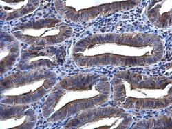
- Experimental details
- SYK antibody [N2C2], Internal detects SYK protein at cytoplasm in human endometrium by immunohistochemical analysis. Sample: Paraffin-embedded human endometrium. SYK antibody [N2C2], Internal (GTX100748) diluted at 1:500.